Introduction
Stomata are small openings that mainly occur on the underside of leaves. They are surrounded by specialised cells and they regulate the gas exchange between the plant and it’s environment, the plant is 'breathing' through them, as it were. Stomata are very recognizable by the two kidney- or bean-shaped guard cells that regulate the size of the opening. The guard cells are specialised epidermal cells which contain vacuoles that change their shape when water is absorbed due to a process called turgor, causing the stomata to open. The stomata are opened by stimuli like high humidity and bright light. Depending on the plant family, guard cells are often surrounded by so-called subsidiary cells.
As for the morphology of stomata, some different shapes can be distinguished:
● anomocytic: without subsidiary cells
● paracytic: with lateral subsidiary cells oriented parallel with the guard cells
● tetracytic: with both lateral and polar subsidiary cells
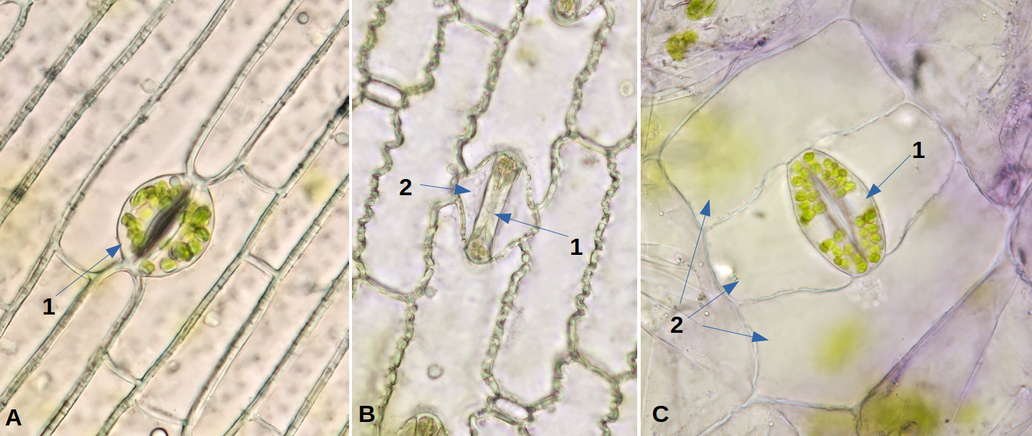
Stomatal shapes in three different monocot plants with indication of guard cells (1) and subsidiary cells (2). A: anomocytic, Dracaena. B: paracytic, maize. C: tetracytic, Tradescantia.
Stomata are fascinating objects to study, in each plant they look a bit different or are positioned differently. To observe stomata we need to peel off the epidermis from the underside of a leaf. If you tear a leaf apart, often a small piece of the epidermis will come off. Especially with thicker leaves this works quite well. Easy to begin with are the leaves of Hosta, Prunus laurocerasus (Cherry laurel) and Tradescantia.
Hosta
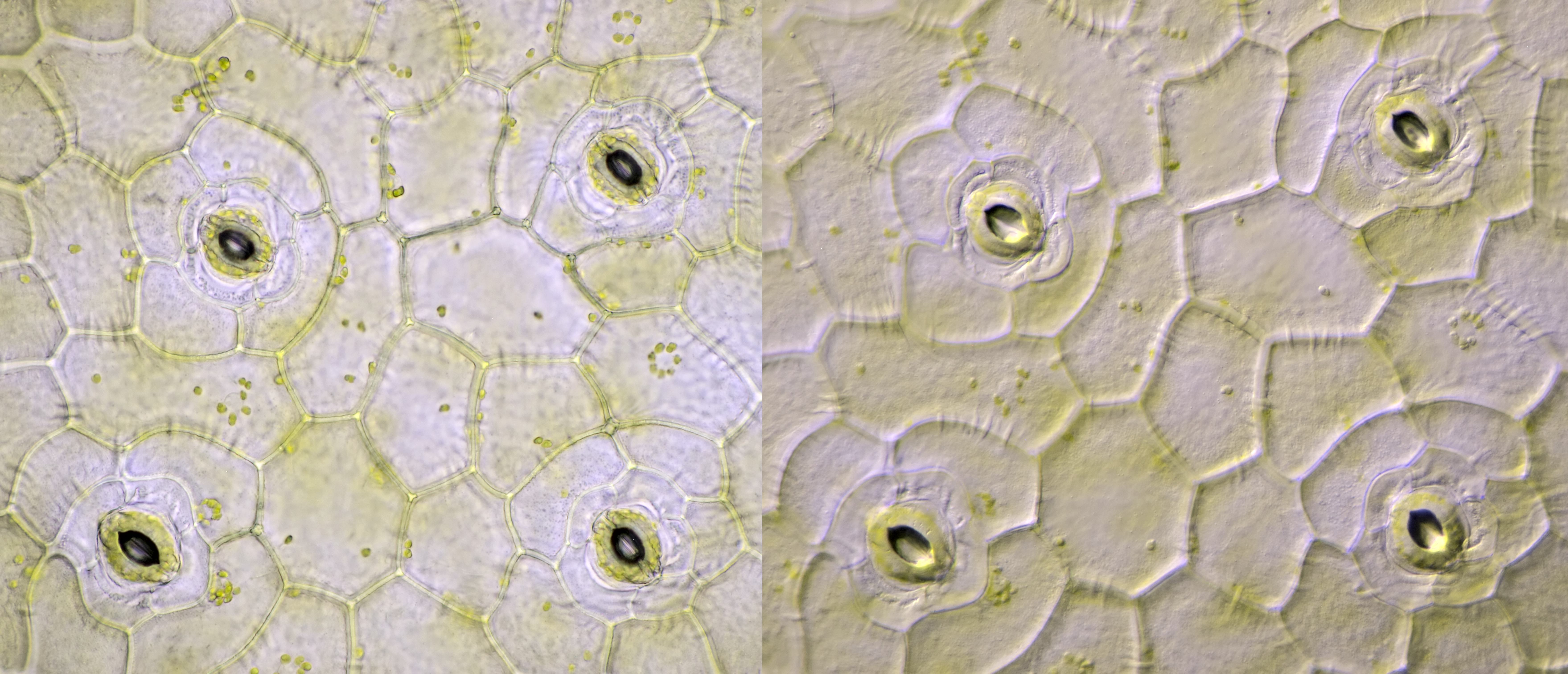
Hostas are popular garden plants. In garden parks you will see them a lot. In the garden park where I have a house, they are also very popular with snails. There are almost no undamaged specimens to be found.
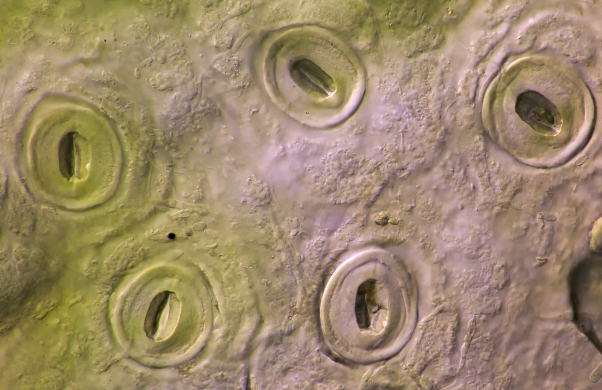
The hosta is a monocotyledonous plant native to East Asia. The epidermal cells are not very typical for a monocot plant and there are no subsidiary cells to be seen. The stomata morphology can therefore be labeled as anomocytic.
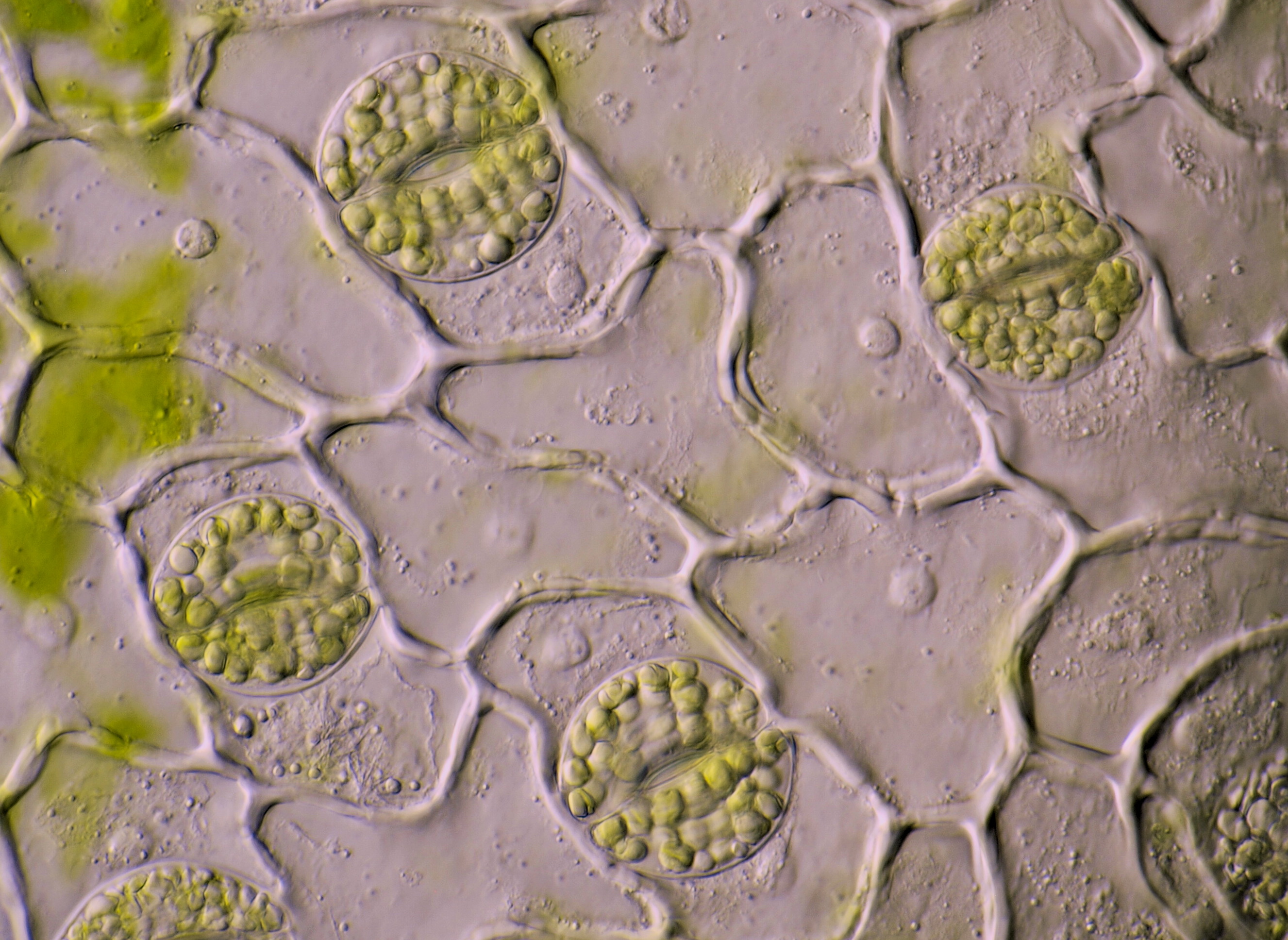
Stomata in a Hosta leaf photographed with oblique illumination. Objective: Carl Zeiss Apo 40/1.0.
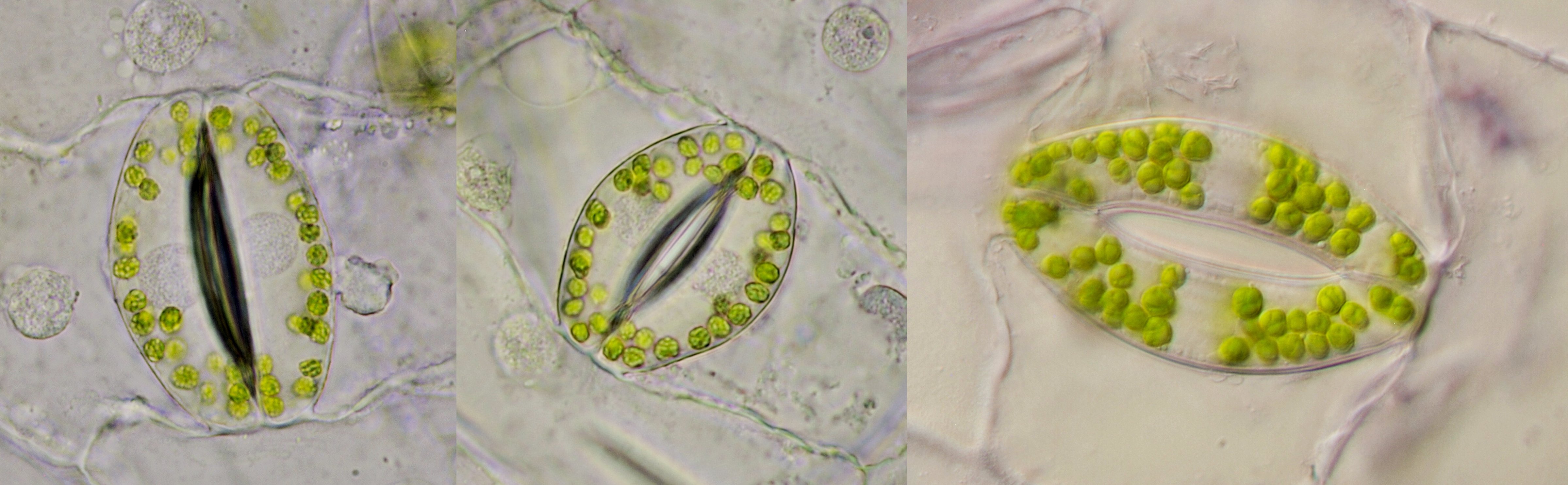
Tradescantia
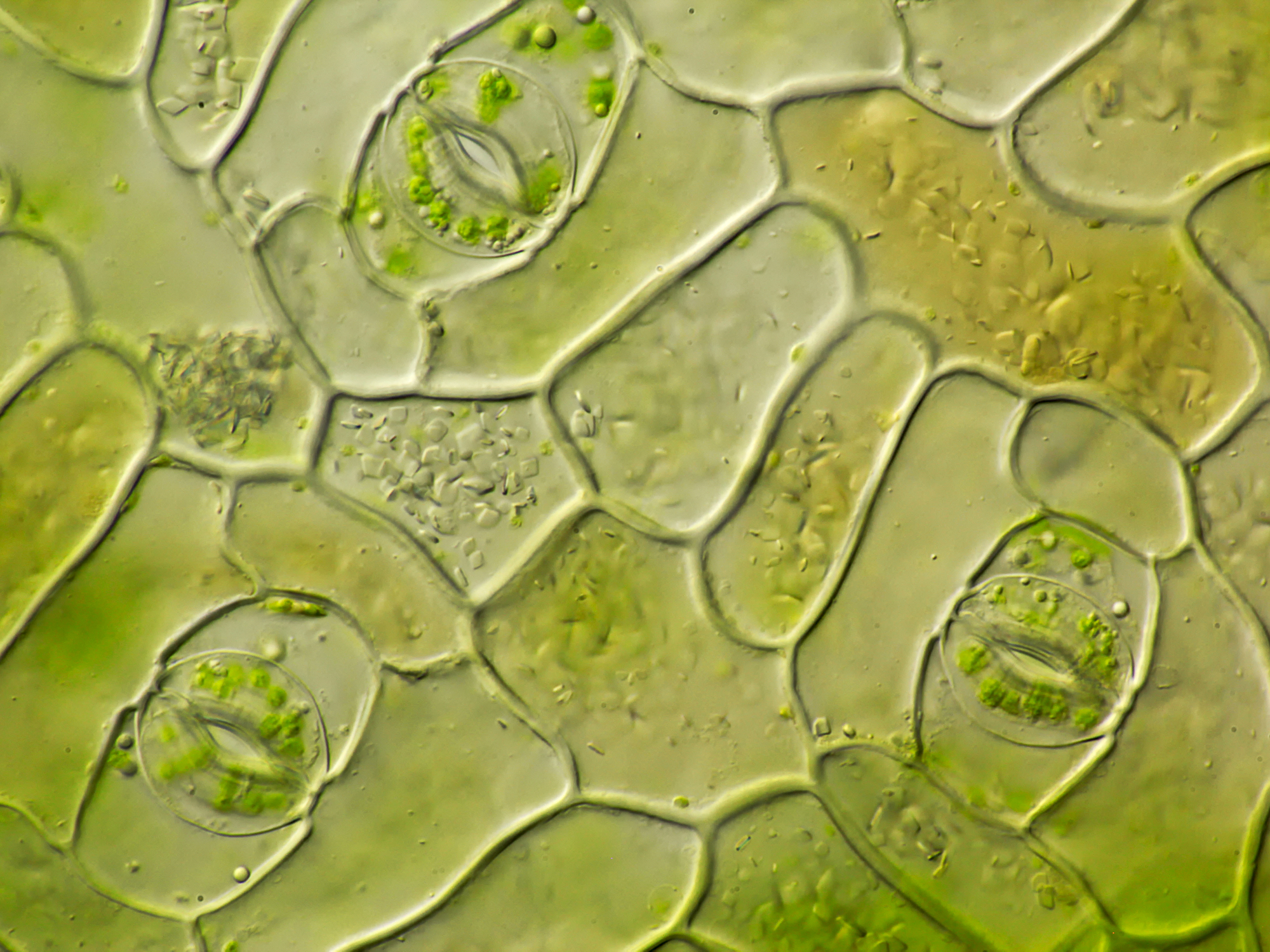
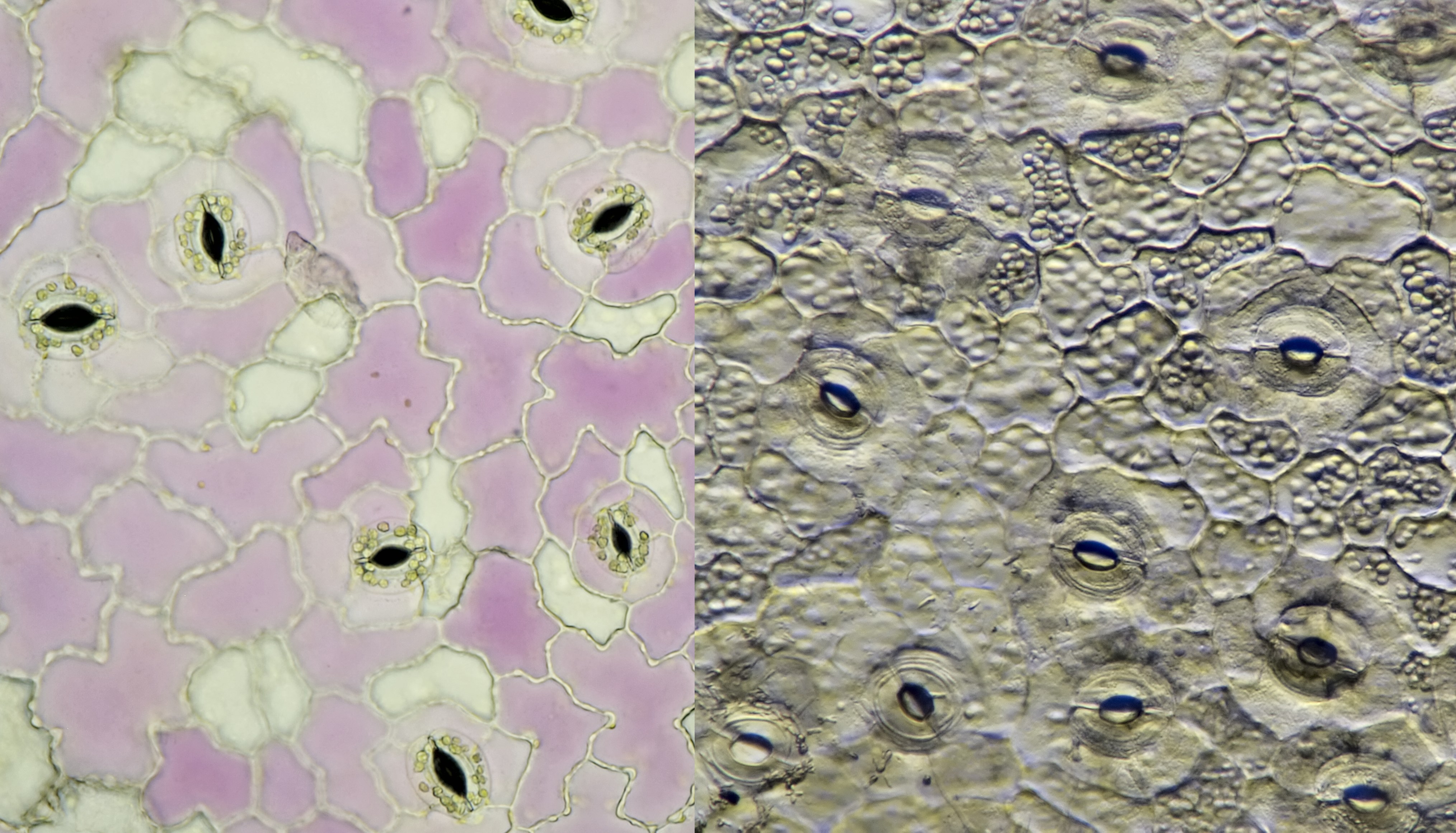
A plant that has always fascinated me is the Tradescantia. This plant has large cells and stomata. When I started with a toy microscope a very long time ago, I observed Tradescantia a lot. At home we had several plants of what was then called Setcreasea purpurea. It’s present name is Tradescantia pallida, a plant native to Mexico. Tradescantia pallida you will find not that often in an average plant store, usually these stores have Tradescantia zebrina, at least in the Netherlands. The latter plant has shorter leaves and a striped pattern. On a microscopical level I could not see any difference between Tradescantia pallida and Tradescantia zebrina.
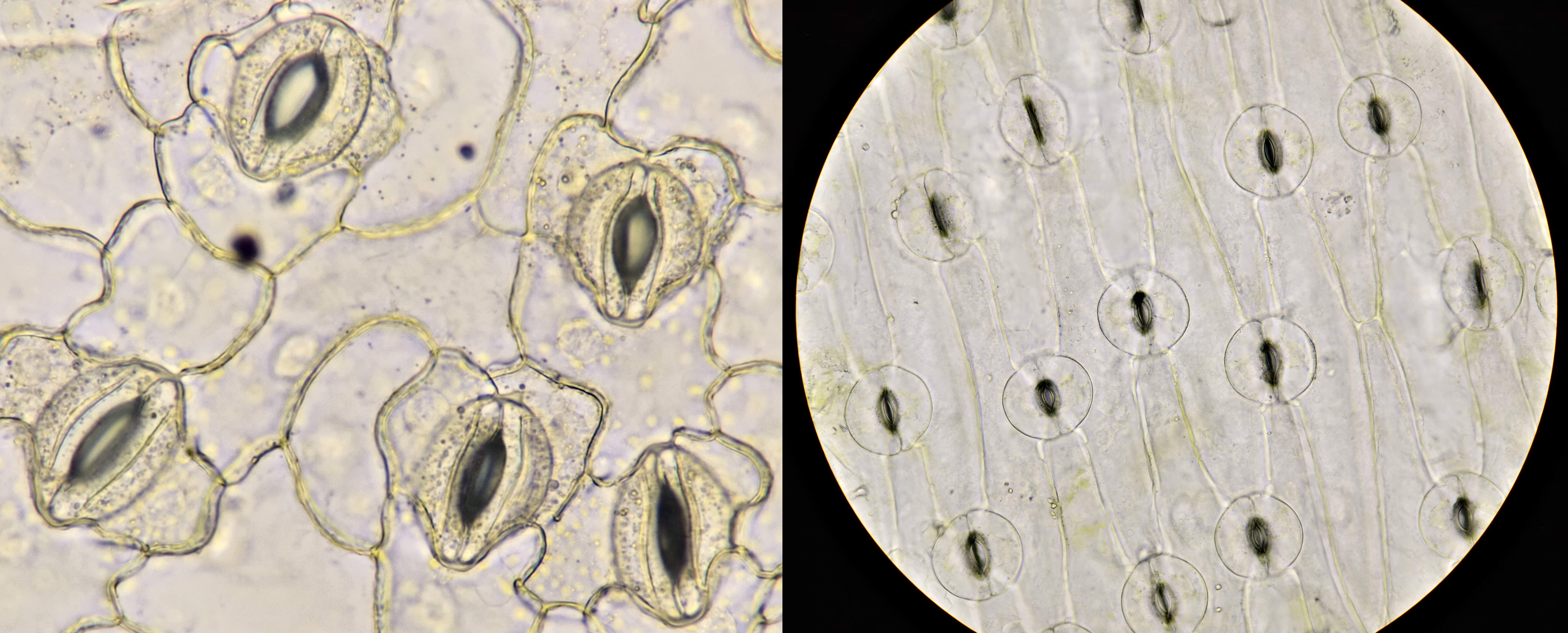
Stomata in the epidermis of Tradescantia zebrina. Objectives: 63/0.85 WI (left), Zeiss-Winkel 40/0.65 (center) en Carl Zeiss Neofluar 63/1.25 (right).

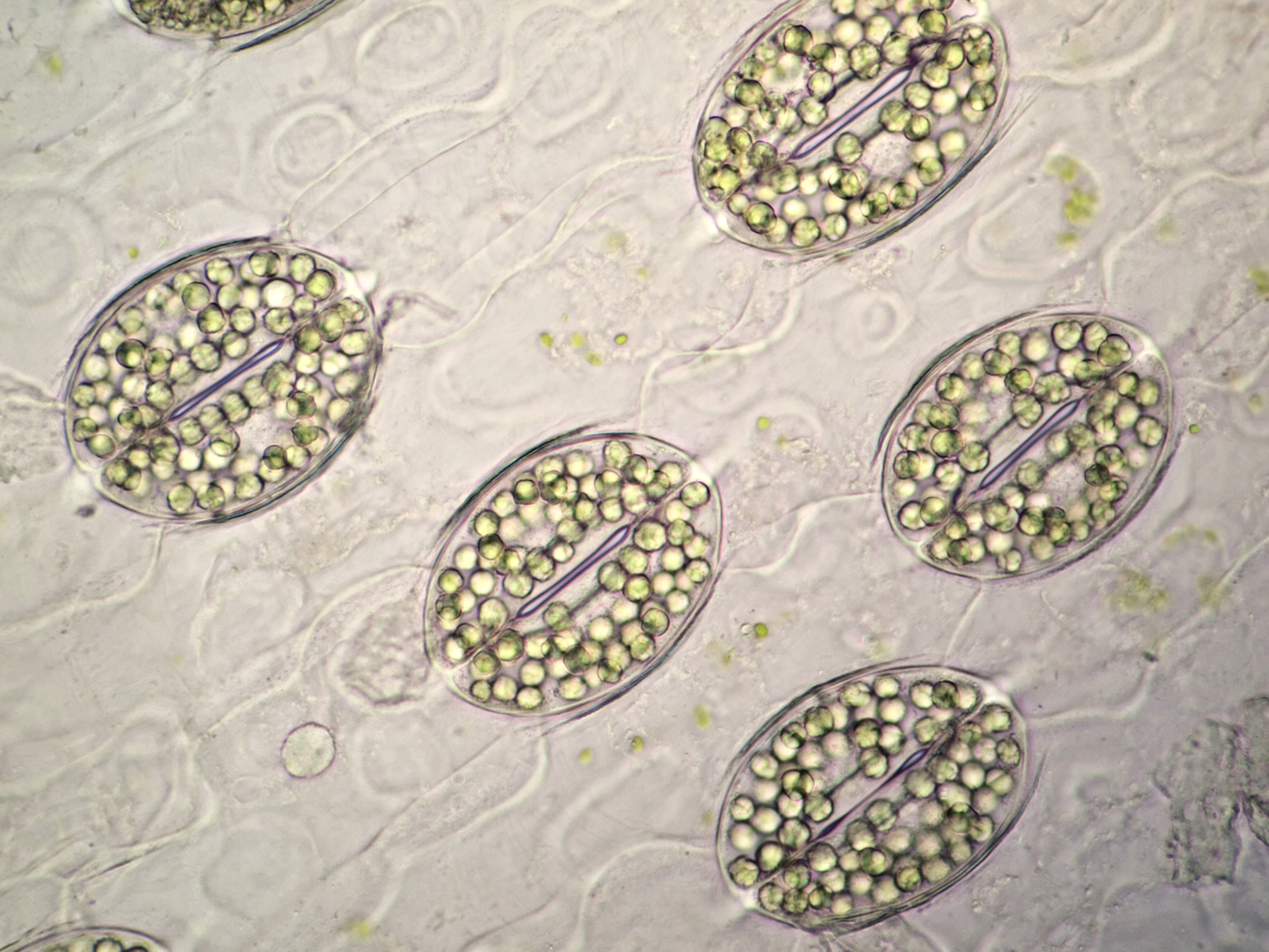
Stomata of Tradescantia zebrina showing the typical arrangement of the subsidiary cells. The nuclei are often surrounded by leucoplasts. Objective: 63/0.85 WI.
These images of Tradescantia zebrina stomata belong to the first photos I ever took of this plant. At that time, as a camera I still used the Olympus Stylus 725 SW, a simple compact camera. Objective: Carl Zeiss 100/1.25.
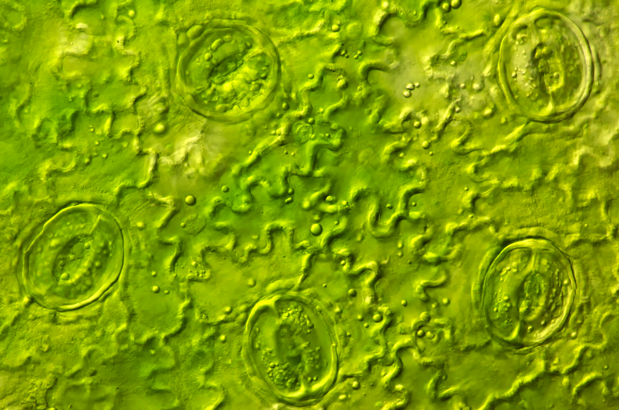
If you look at an intact leaf of Tradescantia zebrina through the microscope, a special picture emerges. The leaves of this plant are quite thick so it’s only possible to use an objective with a long working distance, like a lower magnification objective. Also, a bright light source is needed to shine through the leaf.
A whole leaf of Tradescantia zebrina that was shine through with a bright lightsource. Objective: Zeiss 25/0.45.
Yucca
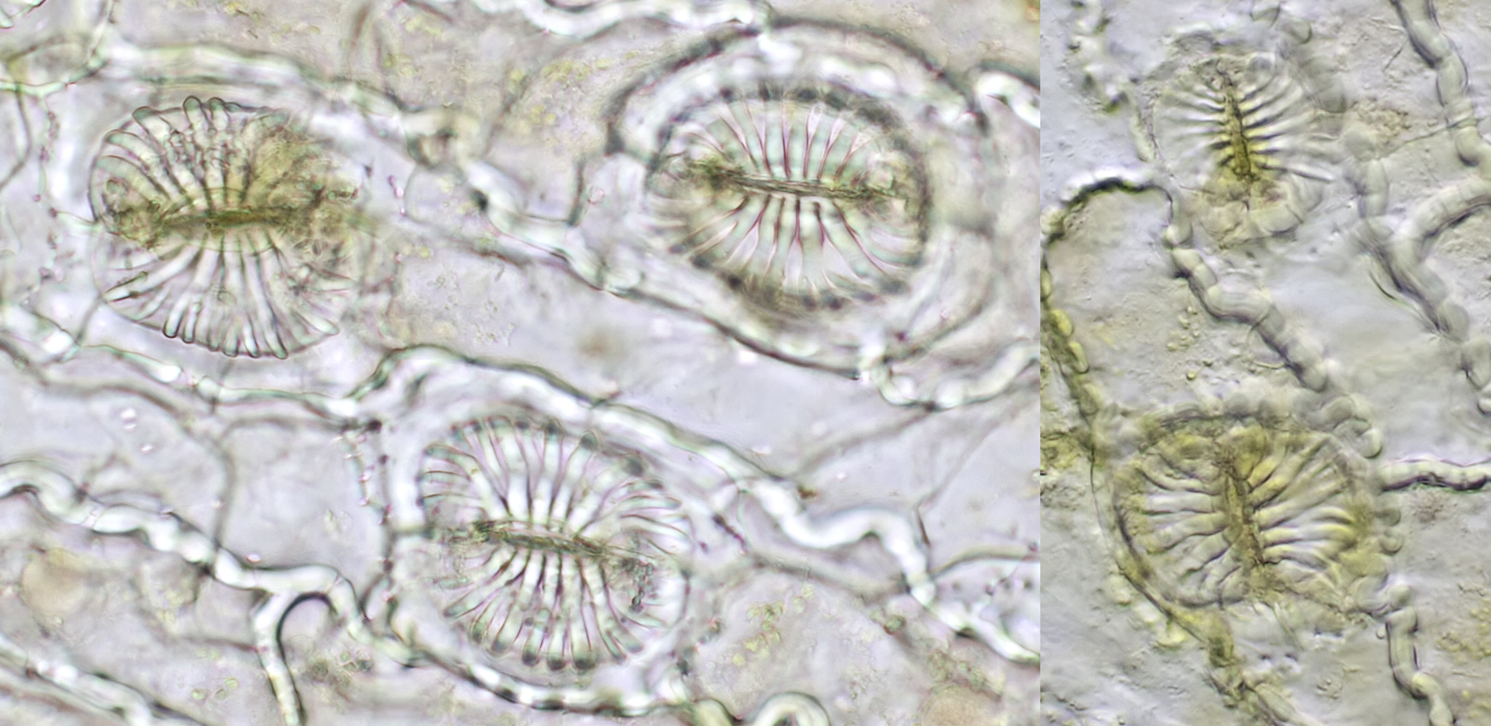
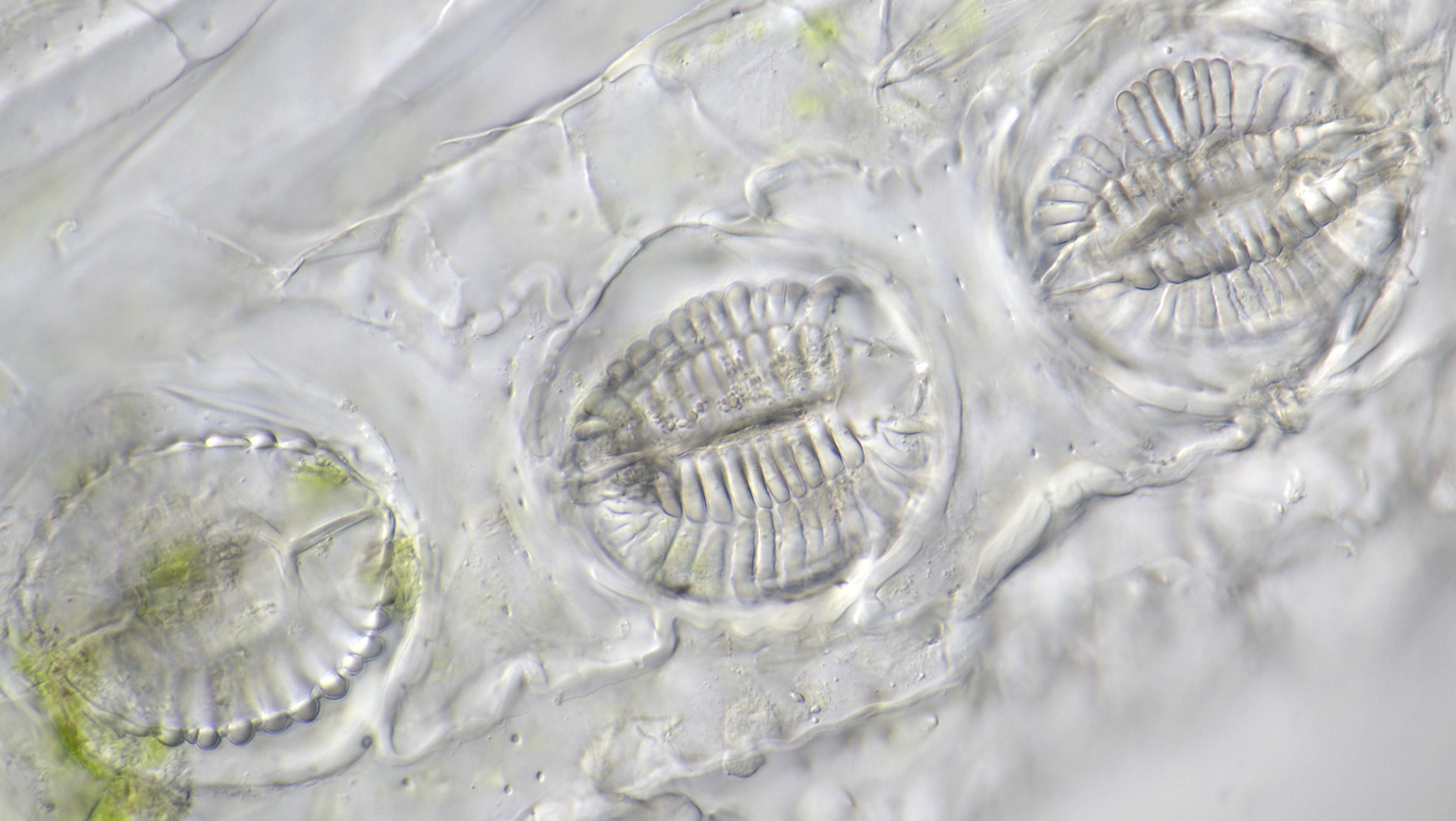
A plant worth investigating is Yucca filamentosa (Palm lily). The stomata of the Palm Lily look quite special. This plant has thick leaves and the epidermis is easy to peel off.
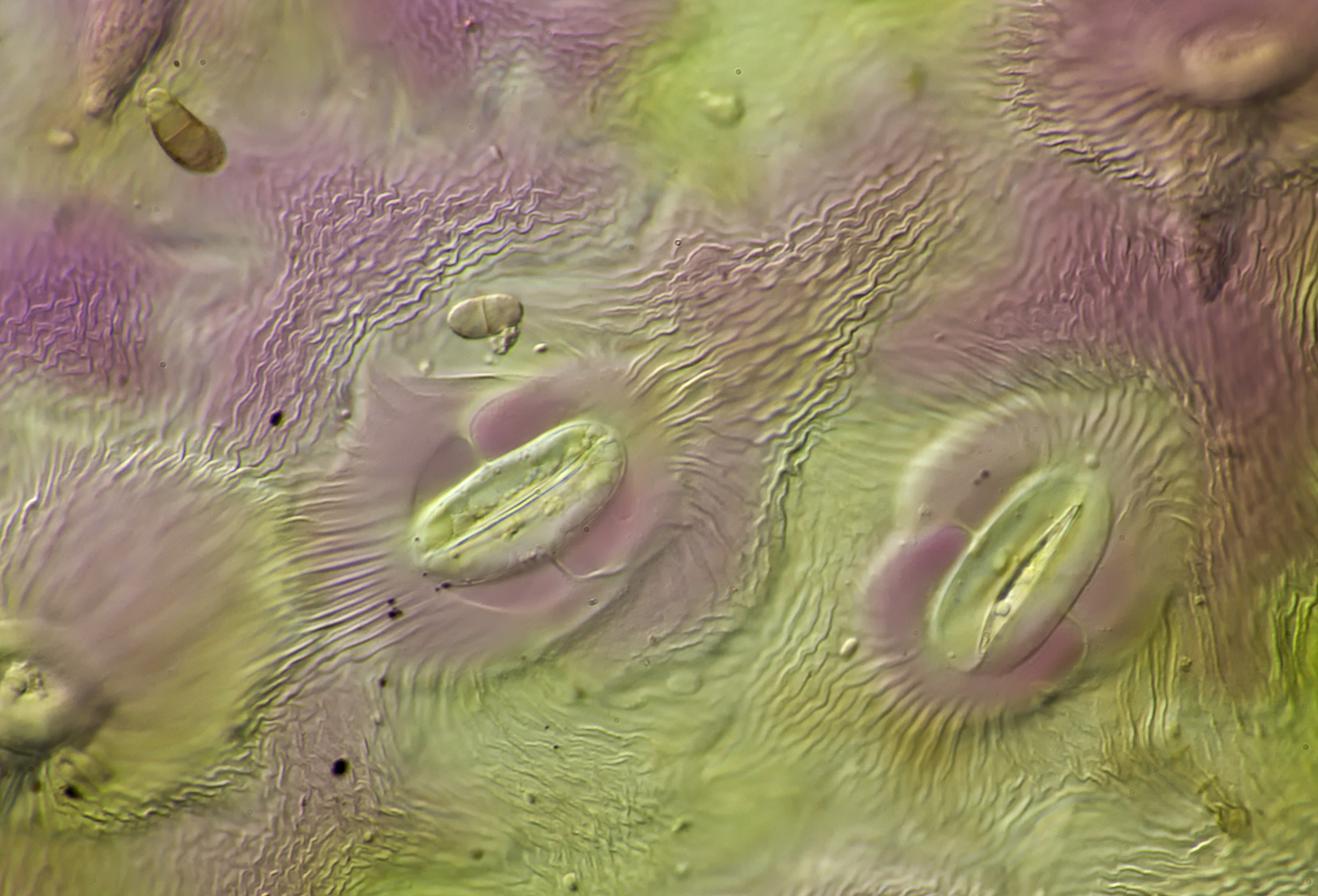
Stomata in the epidermis of Yucca filamentosa. Also visible are many oil droplets. Objective: Neofluar 25/0.60.

Epidermis from Yucca photographed in brightfield illumination (left) and darkfield illumination (right). Objective: Zeiss-Winkel 10/0.25.
The stomata in the petals of Yucca look different from those in the normal leaves. Objective: Zeiss-Winkel 40/0.65.
Maize
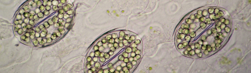
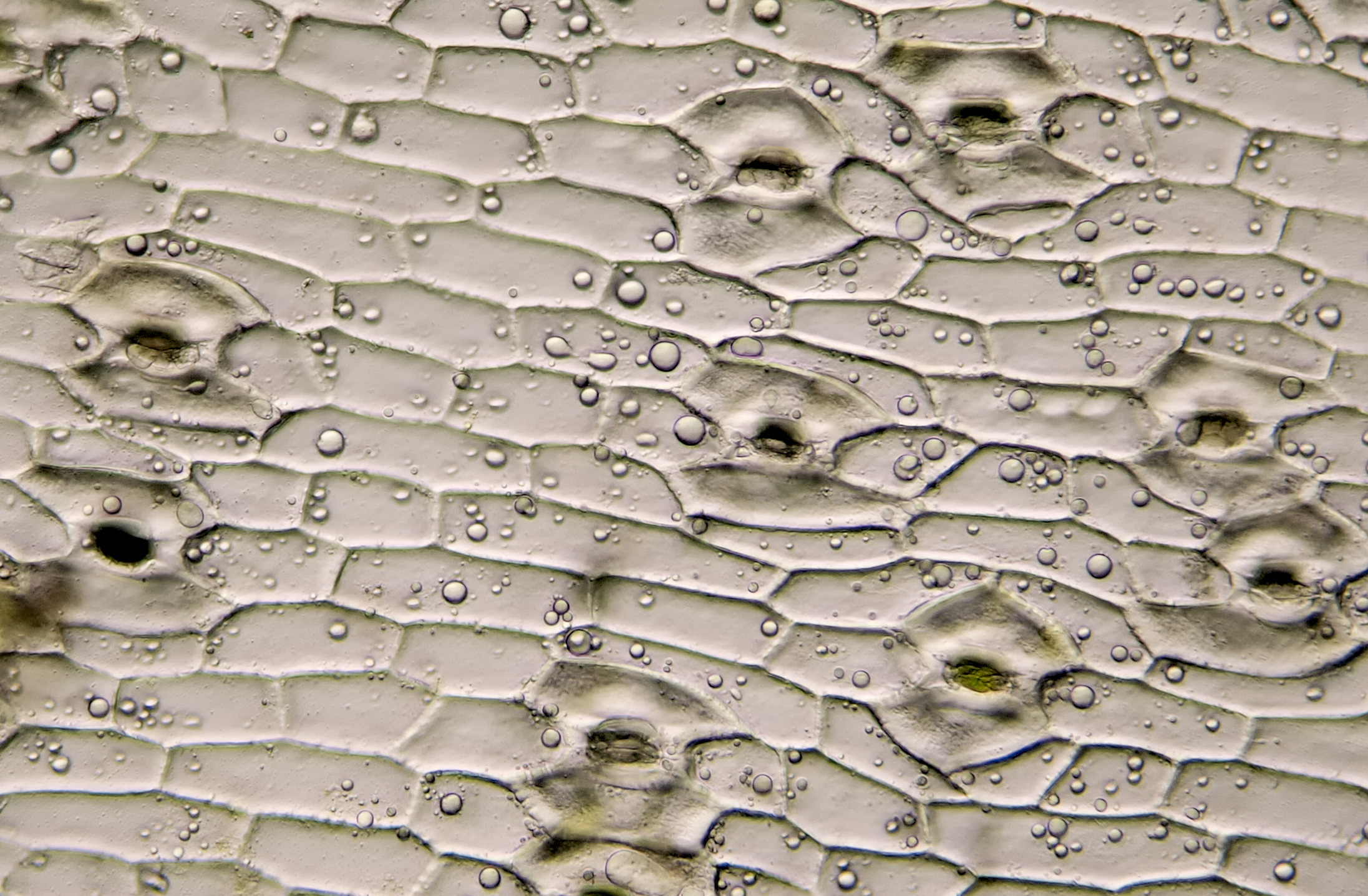
Maize belongs to the grass family (Gramineae) and the epidermis of these plants is characterized by elongated cells with undulating cell walls. The stomata are oriented parallel and have 2 subsidiary cells.
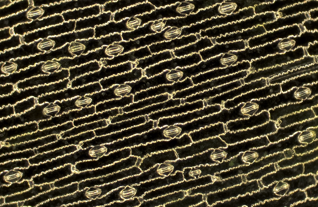
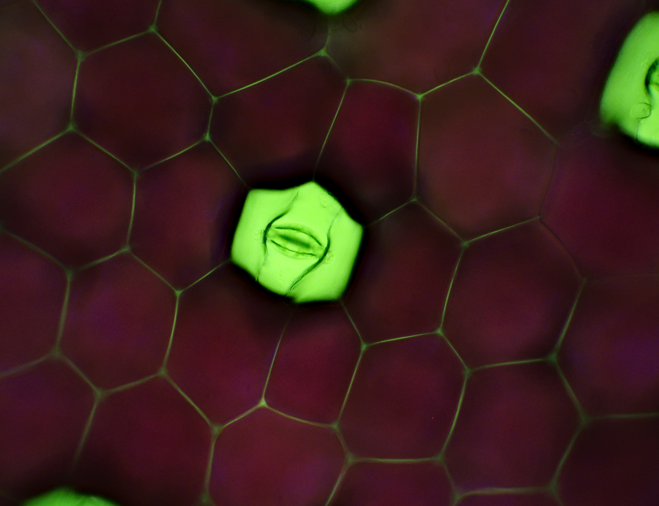
The parallel arrangement of stomata that is often typical of monocotyledonous plants such as in this case, maize. Photographed in darkfield illumination with Zeiss-Winkel 10/0.25 objective.
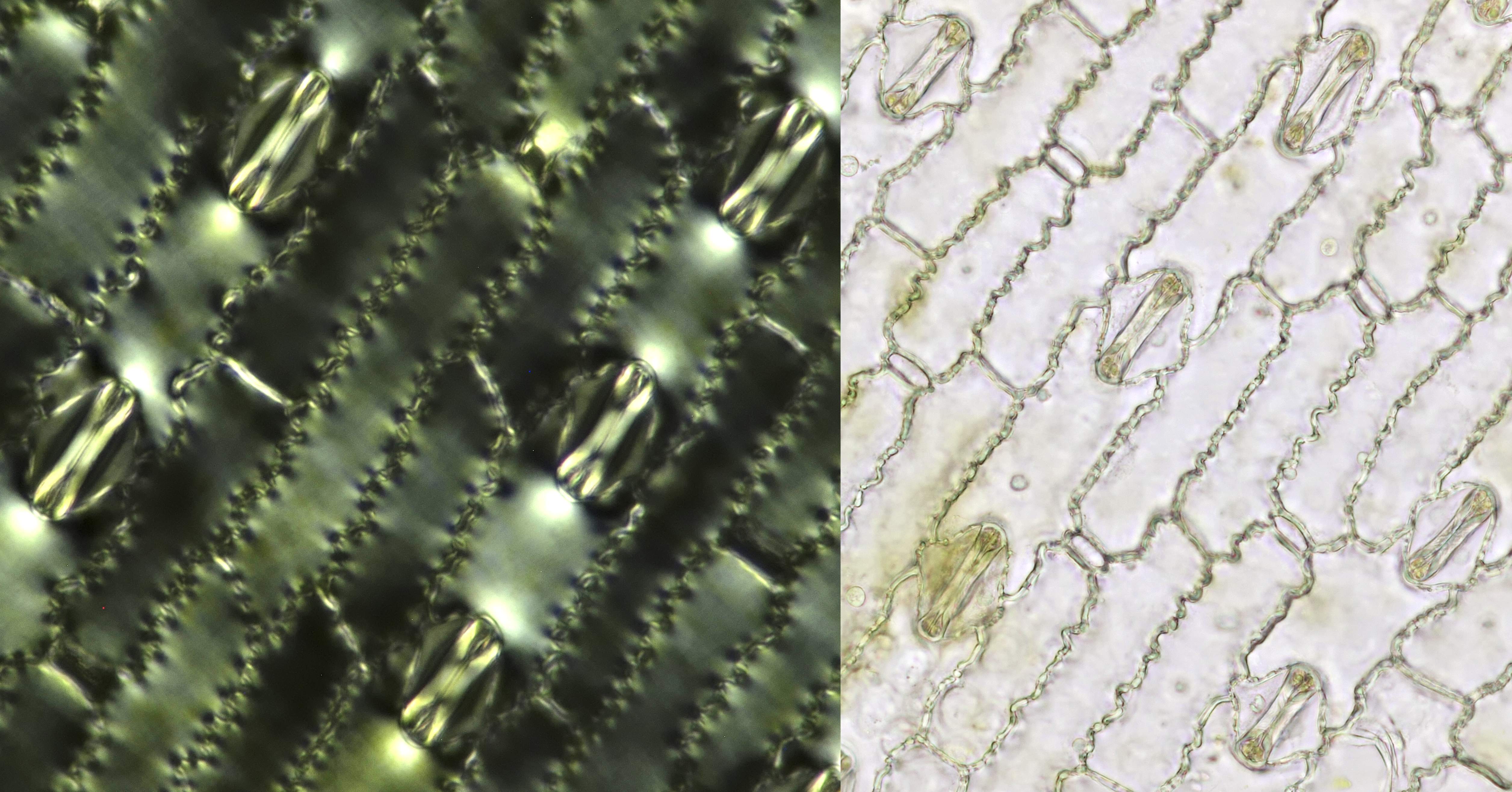
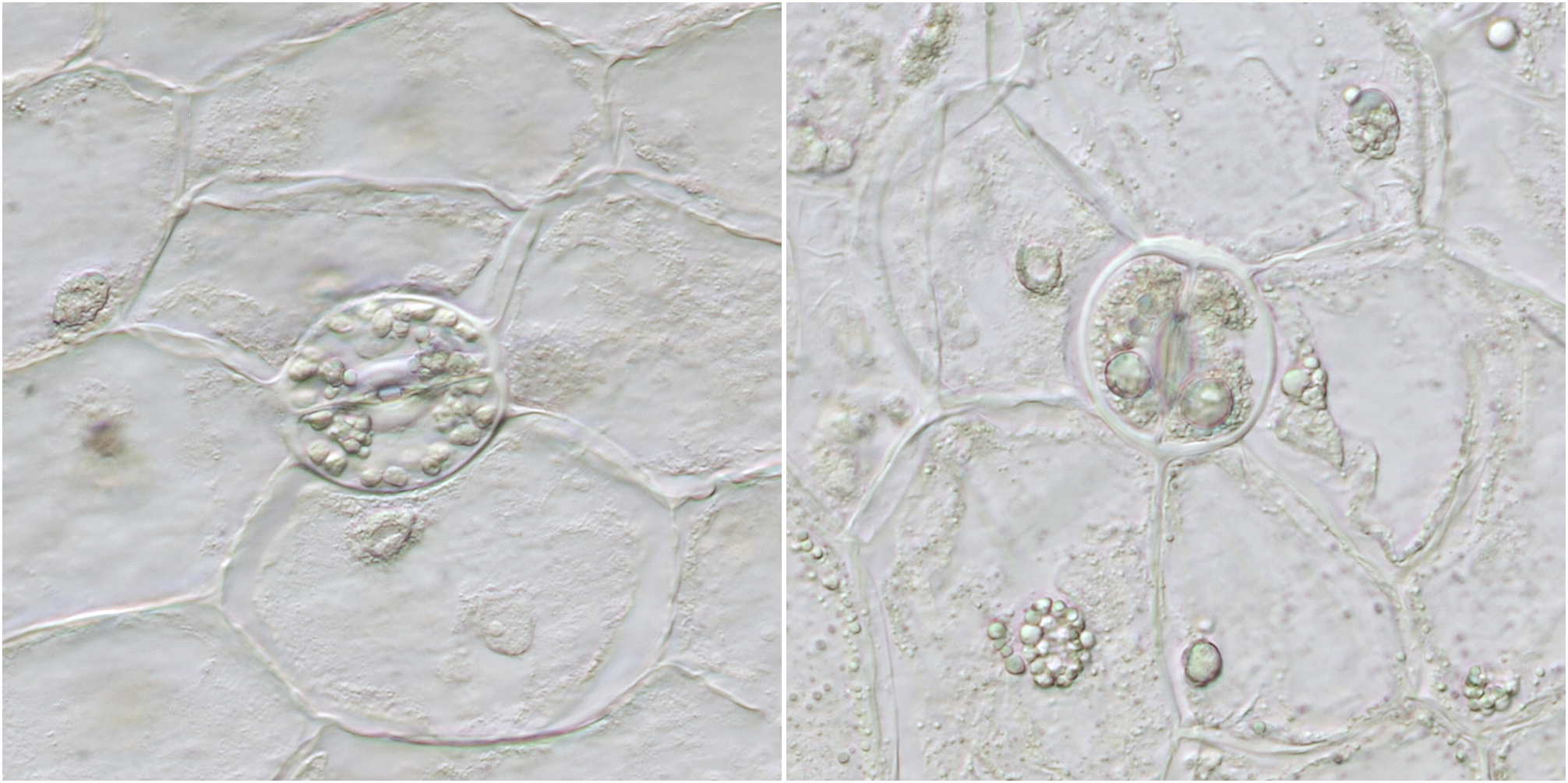
Maize stomata photographed in polarised light (left, objective Zeiss-Winkel 25/0.45) and in brightfield illumination (right, objective Zeiss-Winkel 40/0.65).
Other plants
The following is a selection of stomata in other plants.
Epidermis from a leaf of Canna. In addition to the three stomata, there are many calcium oxalate crystals visible in the cells. Objective: Carl Zeiss Apo 40/1.0.
Stomata in the epidermis of Heldera (Ivy). Objective: Carl Zeiss Apo 40/1.0.
Like the surface of another planet: the epidermis of Acanthus. The patterns are formed by cuticle, a waxy layer that acts as a water repellent. Objective: Carl Zeiss Apo 40/1.0.
Stomata of Prunus laurocerasus (Cherry Laurel). Objective: Carl Zeiss Apo 40/1.0.
Stomata in Brugmansia (Angel's trumpet). Objective: Neofluar 25/0.60.
Left: stomata of Arum (Arum Lily), Olympus objective Plan 20/0.40. Right: stomata in the epidermis of Allium cepa (onion), Zeiss-Winkel objective 40/0.65.
Stomata of Peperomia deppeana photographed in brightfield illumination (left) and oblique illumination (right). Objective: Carl Zeiss Neofluar 40/0.75.
Stomata of Heuchera or coral bells (left, CZJ Apo 16/0.40) and Skimmia (right, Olympus Plan 20/0.40).
Stomata of Equisetum (Horsetail) photographed with Zeiss-Winkel 40/0.65 (left) and Zeiss-Winkel 25/0.45 (right).
Stomata of Equisetum photographed with Carl Zeiss Apo 40/1.0.

A colorful site: the epidermis of purple Basil (Ocimum basilicum) showing anthocyanin-filled vacuoles in guard cells and subsidiary cells. Objective: Carl Zeiss Apo 40/1.0.
Literature
Rudall PJ, Chen ED, Cullen E. Evolution and development of monocot stomata. American Journal of Botany 104 (8): 1122 – 1141 , 2017.
He, Jing Jing & Liang, Yun-Kuan. (2018). Stomata. 10.1002/9780470015902.a0026526.