Depending on the degree of correction, there are roughly three categories of microscope objectives, namely achromates, fluorites and apochromates. Achromats correct the chromatic aberration for 2 colours, fluorites for 3 and apochromates for 4 colours. Apochromats have the highest level of correction and therefore the highest resolution can be achieved with these objectives.
Achromats are often seen as second-rate objectives and less suitable for photography, a common misunderstanding. In my opinion, there is a place for all three categories of objectives and the better corrected objectives cannot always replace the achromats. In many cases, contrast and depth of field will be better with achromats compared to higher corrected lenses. As the numerical aperture (NA) of an objective increases, the higher the resolving power and the shallower the depth of field. A thick slide often does not benefit from the use of a high NA apochromate. I often see images made with apochromats that are disappointing because of the limited depth of field. The use of an achromat in such cases would have resulted in a better impression of the object. It is good to think about which type of objective the object needs. What is important, depth of field or resolving power?
In the following examples I will show a number of comparisons between the different objective classes. It is clear that good photos can be taken with normal achromats. The numbers that are mentioned correspond to the objectives shown in the image at the bottom of this page.
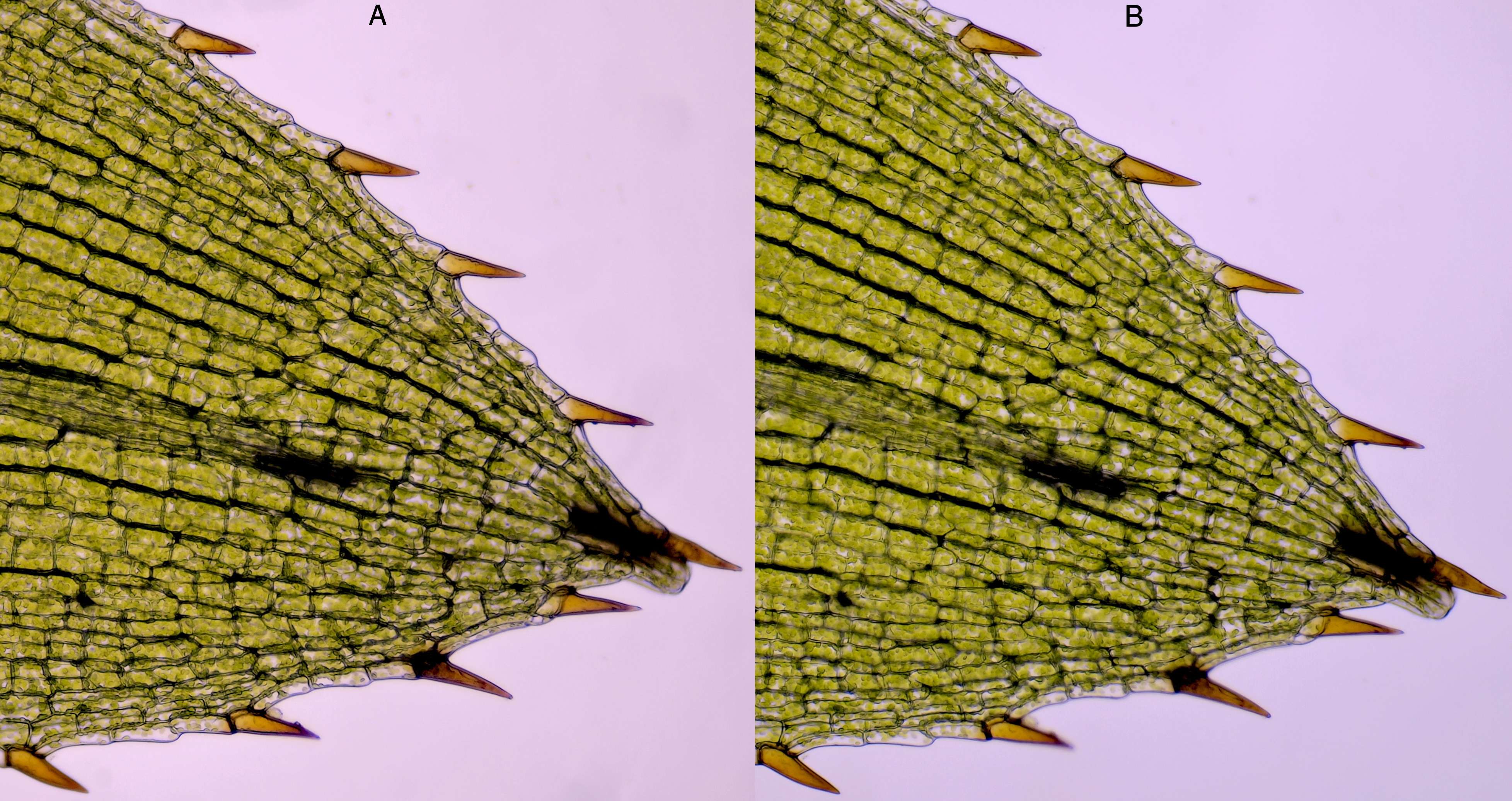
For an overview of a leaf of Ceratophyllum, a Zeiss-Winkel achromat 10/0.25 (A, No. 1) is just as good as a Carl Zeiss Neofluar 10/0.30 (B, No. 2). The achromat gives a little bit more depth of field, which improves the overall impression of this object.

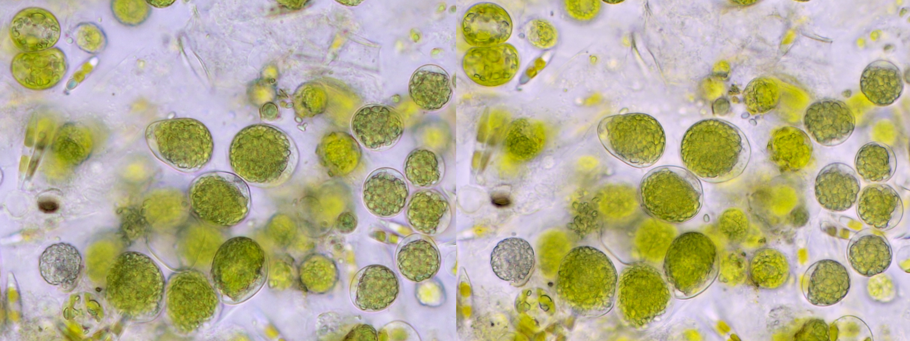
Cosmarium, photographed with a Zeiss-Winkel 40/0.65 achromat (left, No. 3) and a Carl Zeiss 40/1.0 apochromat (right, No. 4).
Arcella, photographed with a Zeiss-Winkel 40/0.65 achromat (left, No. 3) and a Carl Zeiss 40/1.0 apochromat (right, No. 4).
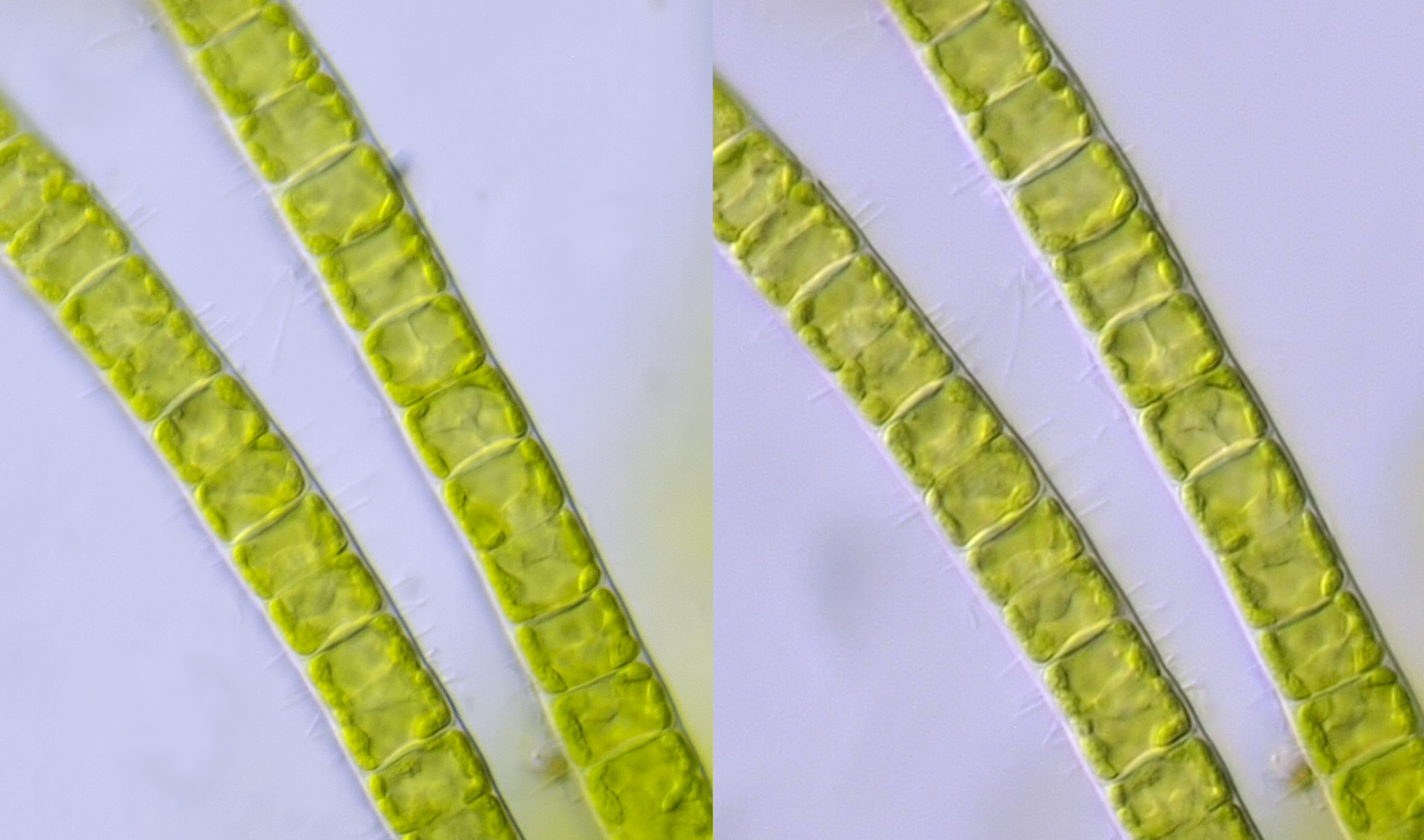
Filamentous algae fotographed with Zeiss-Winkel 40/0.65 achromat (left, No. 3) and Carl Zeiss apochromat 40/1.0 (right, No. 4).
The following comparison is between two completely different objectives: a no-name water immersion achromat 63/0.85 and a Carl Zeiss Neofluar 63/0.9. The 37 mm water immersion objective was attached to an old gray Euromex horseshoe stand. Above all expectations, this objective turns out to be worth it’s weight in gold because water immersion can be applied at a numerical aperture of 0.85. The objective is therefore much less sensitive to deviations in cover glass thickness. The result is an image with high contrast. The Neofluar 63/0.9 objective has a correction collar to adjust for the cover glass thickness. These kinds of dry lenses with high NA are not my favorite, you keep adjusting the cover glass correction all the time and if that is not done properly, the image will be ruined.
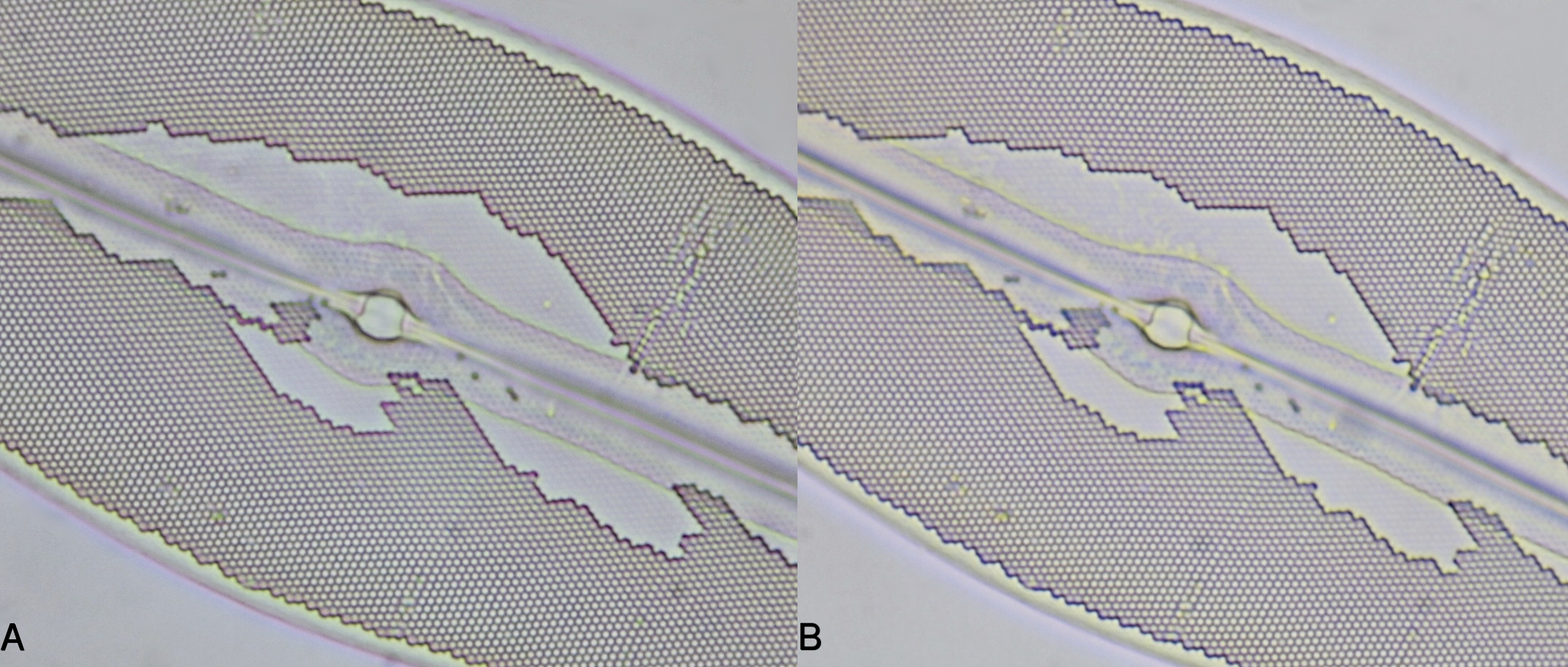
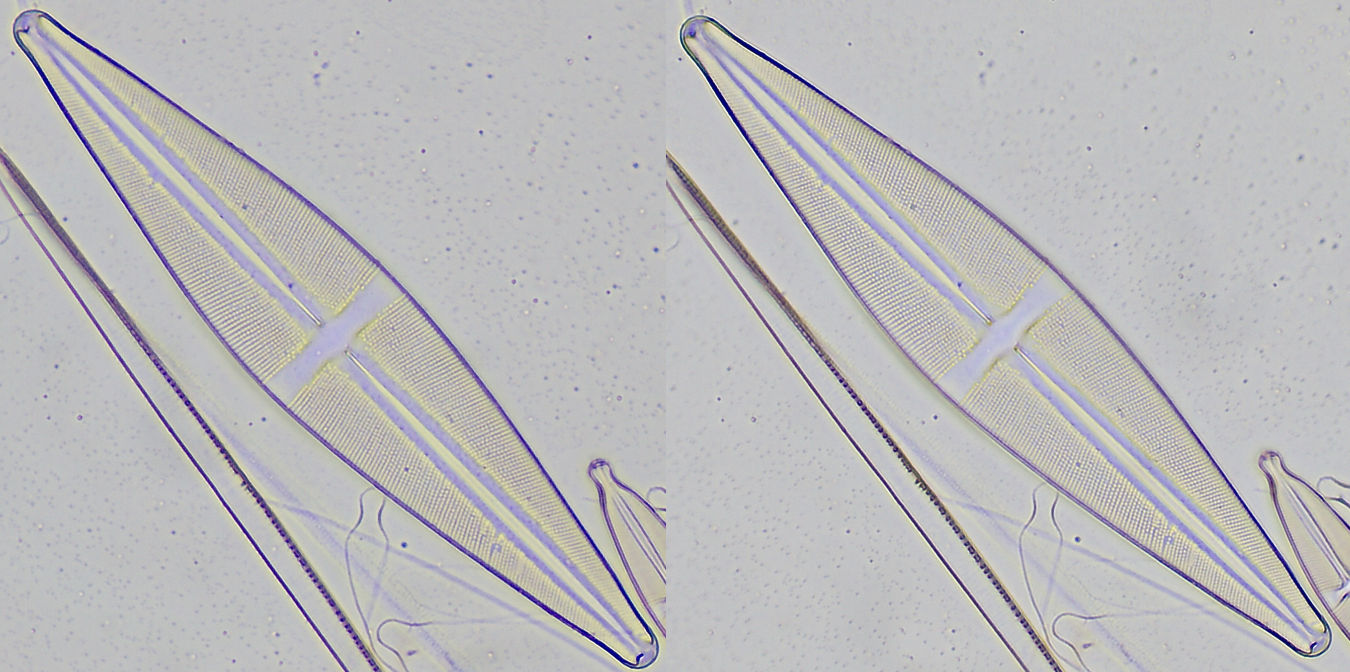
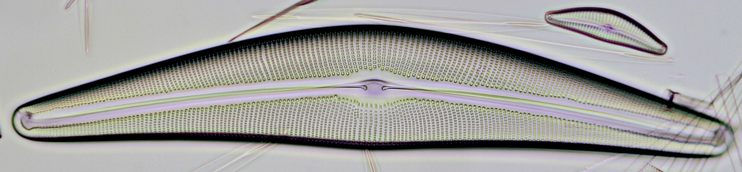
Section of Pleurosigma angulatum photographed with a no-name WI 63/0.85 achromat (A, No. 5) and a Carl Zeiss Neofluar 63/0.90 (B, No. 6). The contrast in image A is slightly better.
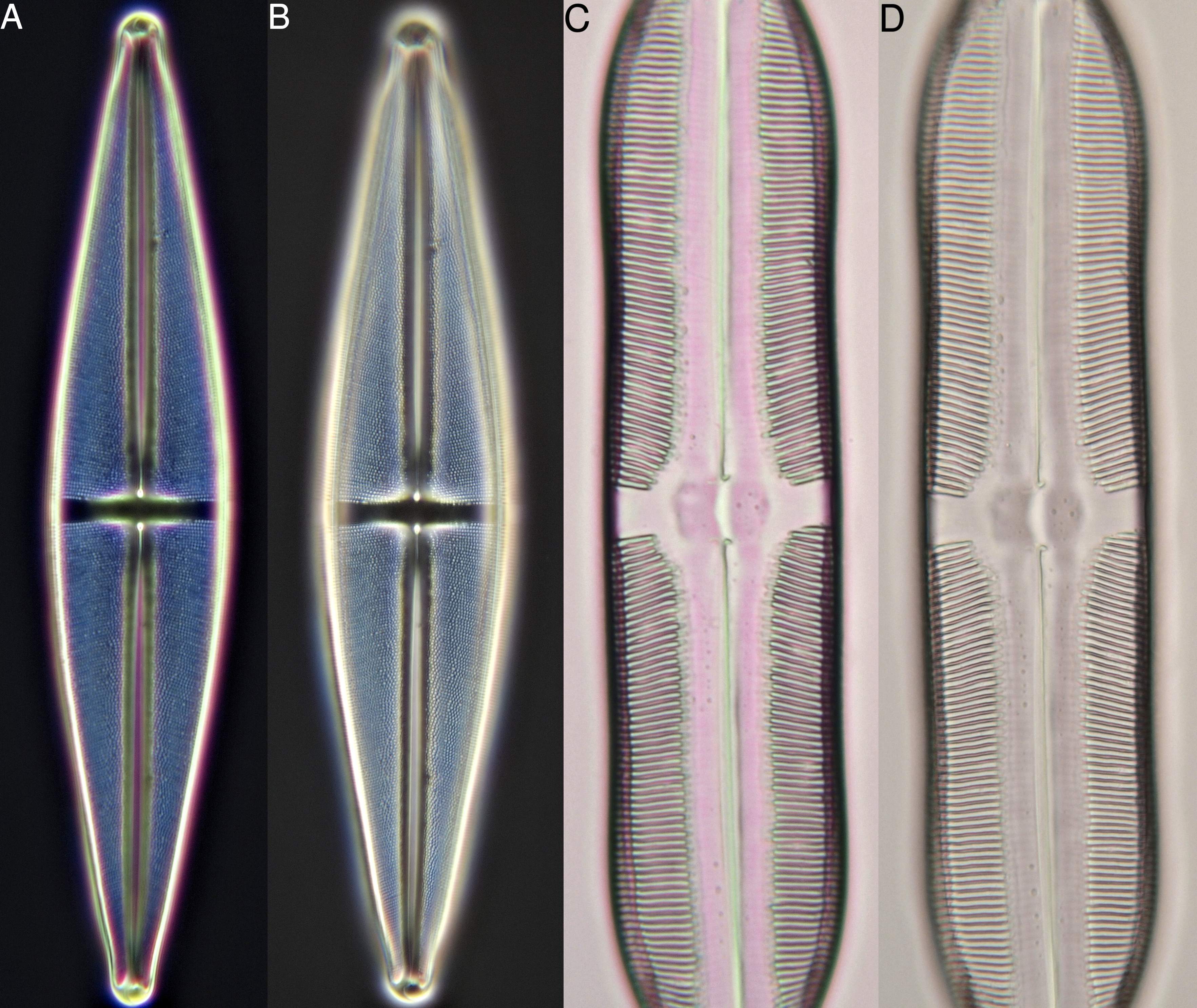
Darkfield image of Stauroneis photographed with a Leitz 25/0.50 achromat (A, No. 7) and a Leitz planapochromat 25/0.65 (B, No. 8). Right: Pinnularia with Leitz achromat 25/0.50 (C) and Leitz planapochromat 25/0.65 (D). More chromatic aberration is visible with the achromat, but the depth of field is better. There is also more glare visible in the darkfield image from the planapochromat; a higher aperture often gives lesser results in dark field.

Mix of different types of pollen grains photographed with a Leitz 25/0.50 achromat (left, No. 7) and a Leitz planapochromat 25/0.65 (right, No. 8). The image on the right is of better quality, but it is debatable which image gives the best subjective impression. Due to the better depth of field with the achromat, the boundaries of the pollen grains are clearer.

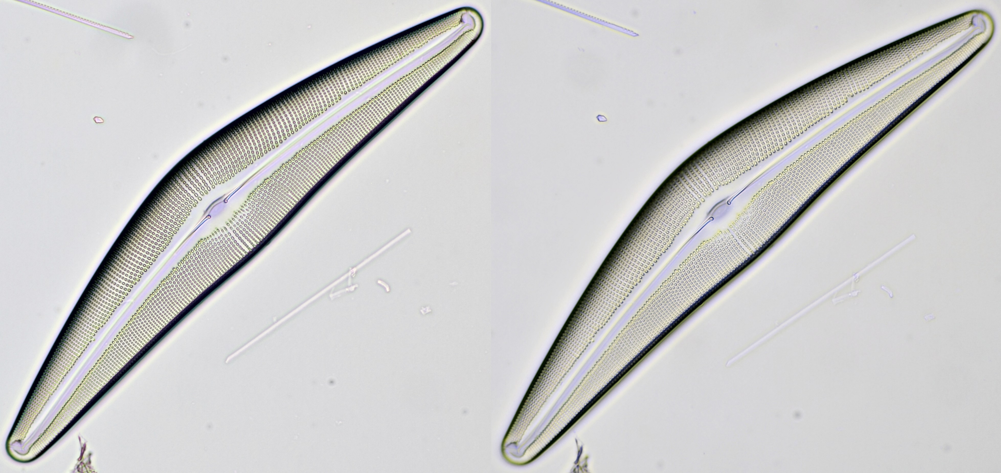
Darkfield images of Pleurosigma angulatum. Leitz 25/0.50 (upper image, No. 7) and Leitz Planapo 25/0.65 (lower image, No. 8). The blue color in the upper image is caused by interference and is only visible when using the achromat. I must say that I find this blue colour quite pleasant to look at in darkfield. The structure is of course better resolved with the planapochromat.
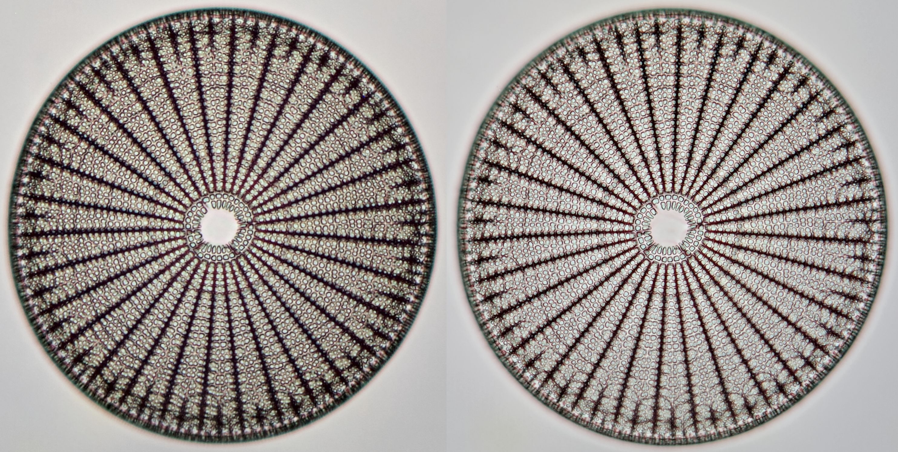
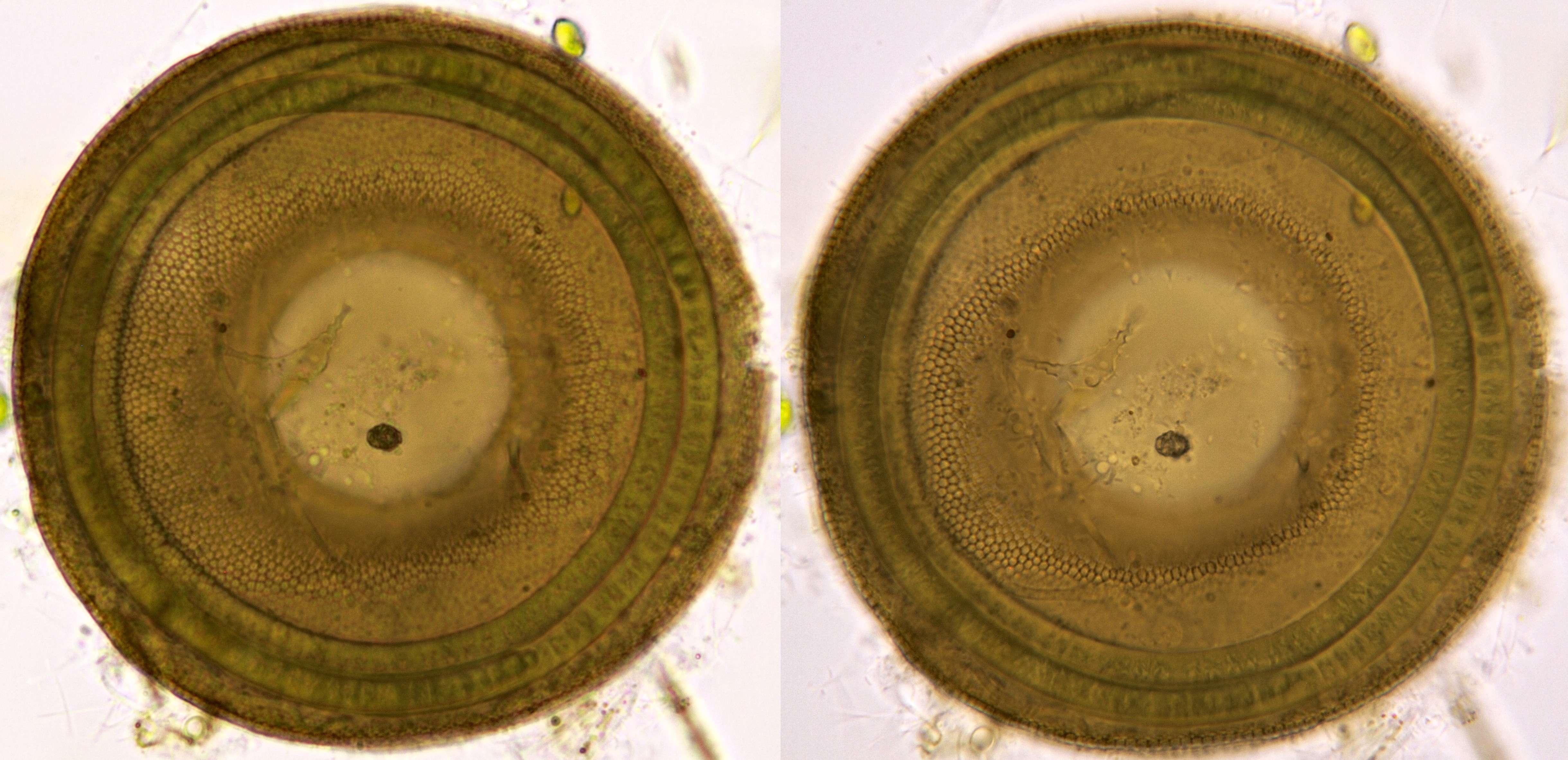
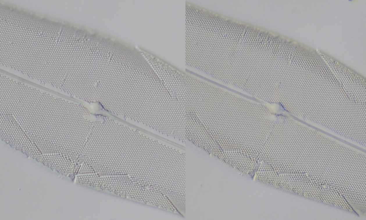
Arachnoidiscus ornatus photographed with Zeiss-Winkel 25/0.45 (left, No. 9) and Carl Zeiss Neofluar 25/0.60 (right, No. 10). The difference in resolution is visible: details are sharper with the Neofluar.

Arachnoidiscus ornatus photographed with Leitz EF 25/0.50 (left) and Leitz NPL Fluotar 25/0.55 (right). The difference here is very subtle and not as clear as in de previous image, also because of the minimal difference in aperture between te two objectives. The image from the EF 25/0.50 has more contrast.
A moss, photographed with circular oblique illumination coming from the Heine condenser. A: Leitz 25/0.50 (No. 7). B: Leitz Planapo 25/0.65 (No. 8). Is it clear that the achromat can handle this kind of illumination in this specimen much better than the planapochromat, which gives a disturbing image.
Cosmarium photographed with Leitz achromat 25/0.50 (left, No. 7) and Leitz Pl Apo 25/0.65 (right, No. 8).
Comparison between Leitz 25/0.50 (left, No. 7) and Leitz Pl Apo 25/0.65 (right, No. 8) in normal brightfield illumination. The structures of the diatoms are better resolved with the planapo while with the achromat more contrast and depth of field is seen.
The same slide as in the previous image, now photographed in darkfield illumination created with the Heine condenser. Leitz 25/0.50 (left, No. 7) and Leitz Pl Apo 25/0.65 (right. No. 8). Some details are more clear with the achromat because of better depth of field.
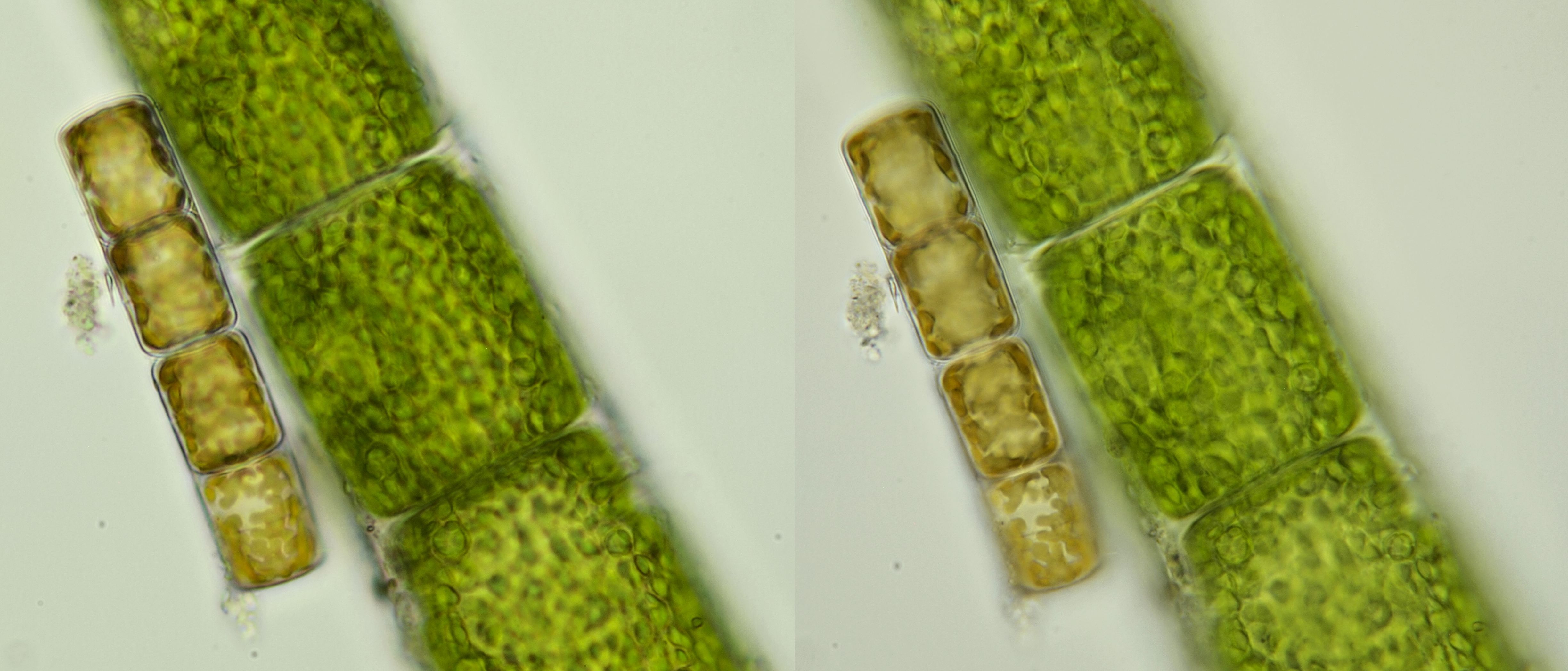
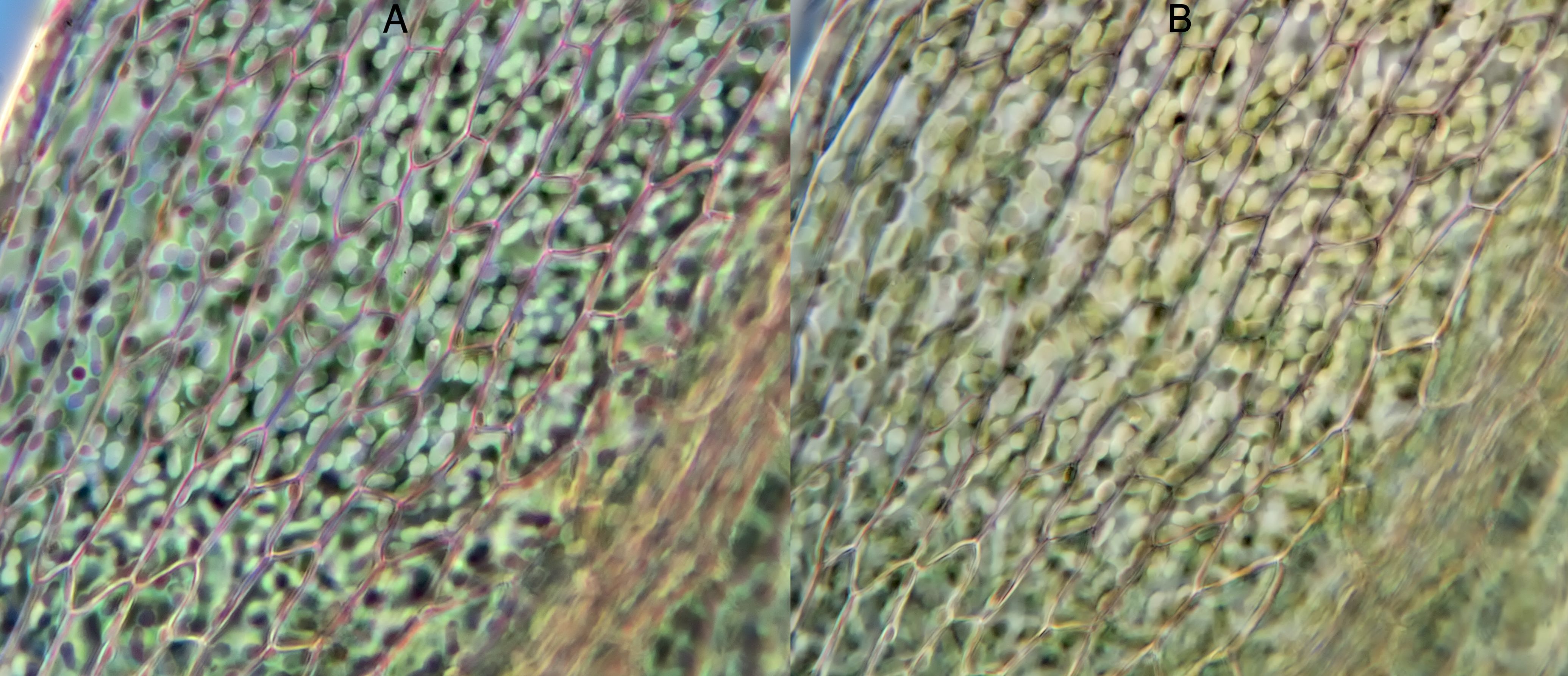
Cladophora, covered with various cyanobacteria. Left: Leitz 25/0.50. Right: Leitz Pl Apo 25/0.65. The image on the left gives a better overall impression.
Left two images: Oedogonium photographed with Zeiss-Winkel achromat 40/0.65 (No. 13) and Carl Zeiss Neofluar 40/0.75 (No. 12). Right two images: Pinnularia photographed with Zeiss-Winkel 40/0.65 (No. 13) and Carl Zeiss Neofluar 40/0.75 (No. 12).
Stauroneis, photographed with a Carl Zeiss achromat 40/0.65 (left, No. 11) and Carl Zeiss Neofluar 40/0.75 (right, No. 12).
Cymbella, photographed with Zeiss-Winkel achromat 40/0.65 (left, No. 3) and Carl Zeiss Neofluar 40/0.75 (right, No. 12). The details are more clear with the achromat when viewing the image at a smaller size.
Mix of different algae photographed with a Zeiss-Winkel 40/0.65 achromat (left, No. 13) and a Carl Zeiss Neofluar 40/0.75 (right, No. 12). The colours are better with the Neofluar while the achromat gives a better depth of field.
Pleurosigma angulatum, photographed with oblique illumination. Left: Zeiss-Winkel 40/0.65 (No. 13). Right: Carl Zeiss Neofluar 40/0.75 (No. 12). The resolution and colour correction in both images is comparable. Personally, I find the visibility of the structure slightly better with the achromat.
Filamentous algae photographed in oblique lighting with Carl Zeiss Apo 40/1.0 (left) and Zeiss-Winkel 40/0.65 (right). On the right there is clearly more depth of field which leads to a better impression of the object.
The following examples show that decent images can be made with simple achromats.
Cymbella, photographed with Zeiss-Winkel achromat 40/0.65 (No. 3).
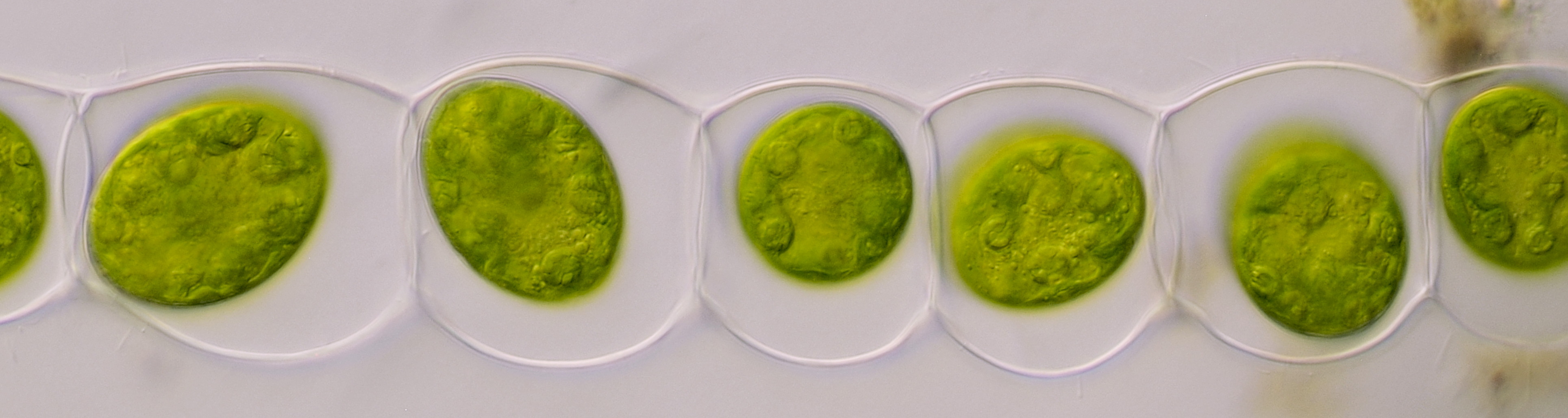
Zygotes of Spirogyra. Zeiss-Winkel achromat 40/0.65 (No. 3).
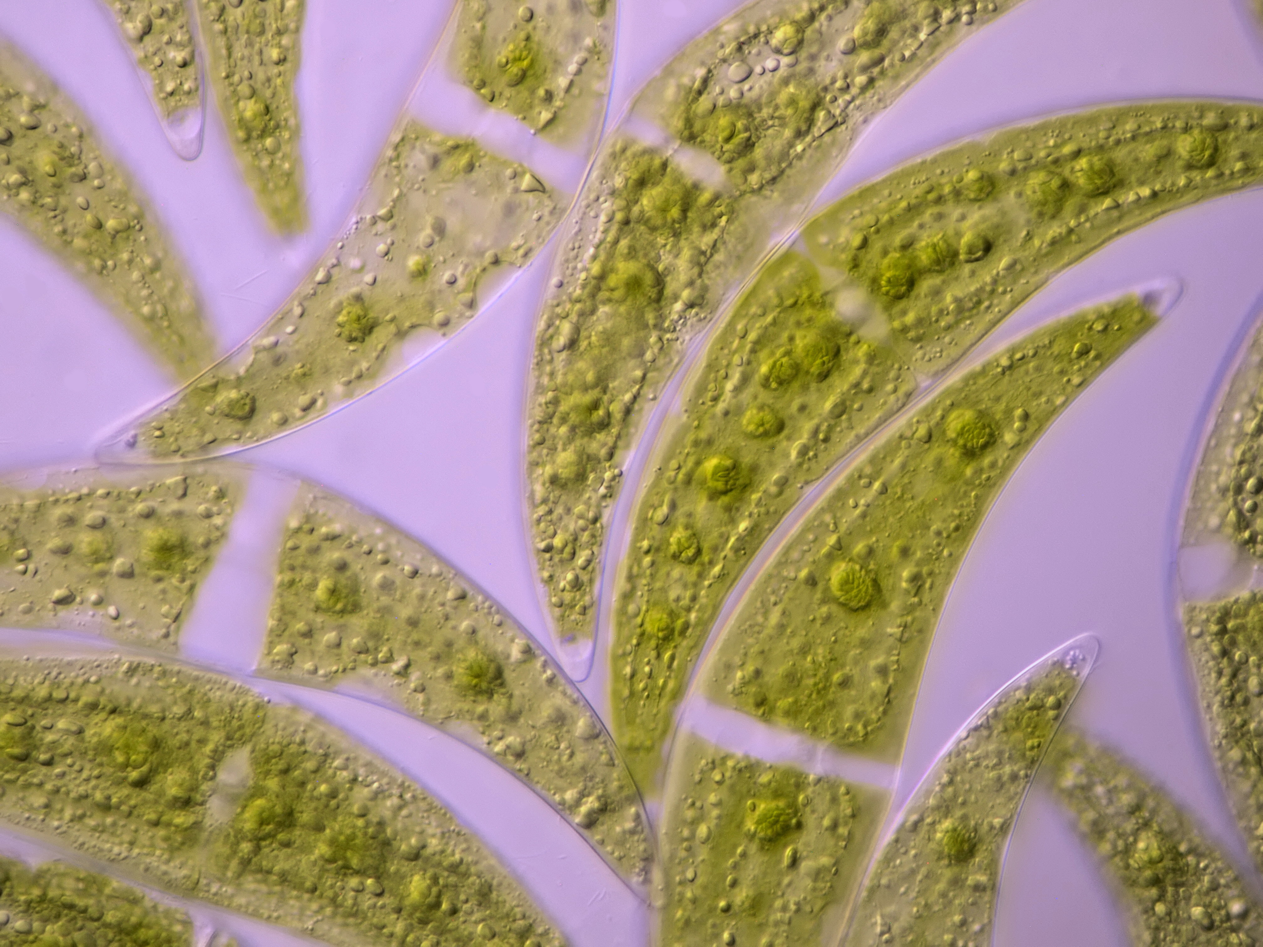
Closterium, photographed with oblique illumination. Carl Zeiss achromat 40/0.65 (No. 11).
The objectives
The objectives used for these tests are shown below. The numbers are indicated with the previous images.
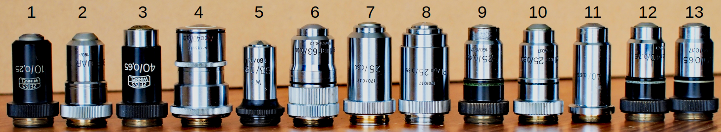
1: Zeiss-Winkel 10/0.25
2: Carl Zeiss Neofluar 10/0.30
3: Zeiss-Winkel 40/0.65
4: Carl Zeiss Apo 40/1.0
5: WI 63 / 0.85, no-name
6: Carl Zeiss Neofluar 63/0.9
7: Leitz 25/0.50
8: Leitz Pl Apo 25/0.65
9: Zeiss-Winkel 25/0.45
10: Carl Zeiss Neofluar 25/0.60
11: Carl Zeiss 40/0.65
12: Carl Zeiss Neofluar 40/0.75
13: Zeiss-Winkel 40/0.65
Conclusion
Achromats should deserve a little more appreciation. These objectives are good all-rounders and they are also fully usable for photomicrography. I think that better corrected objectives are chosen way too often for visual observation and photography. This is not necessary. In fact, in some cases the image from an apochromat will even be disappointing because the object to be examined needs more depth of field. Resolution comes at the expense of depth of field. And many objects lack the fine details that justify the use of a high NA apochromat. Achromats are simply build and contain fewer lenses than, for example, (plan) apochromats, which translates into a better contrast.
There will be microscopists who only use the highest corrected objectives and who consider the achromats to be inferior. However, the expensive high quality objectives they use are no guarantee for good images.
It can be an eye-opener to show micro photos to people who have no idea about microscopy. These people look at these images with completely different eyes than an experienced microscopist. My experience is that, in comparison tests, the photos taken with achromats are usually picked out as being the best because of better depth of field which gives objects clearer boundaries and thus better overall impression.