With normal bright field illumination, a specimen is evenly illuminated from all sides. When the light that illuminates the specimen is coming from one side, it is called oblique illumination. With oblique illumination, less light is shining directly through the specimen. In stead, the light rays are illuminating the specimen at an angle which gives a relief effect. This illumination technique is very old and is used to increase the visibility of difficult specimens.
With oblique illumination, both contrast and the visibility of fine details are enhanced. A pseudo 3-dimensional effect is created that gives depth to structures in the specimen thereby increasing visibility.
Oblique illumination can be achieved in many different ways. By simply holding a finger between the light source and condenser, already a kind of oblique lighting is achieved. In principle, any intervention in the light path that results in an uneven illumination of the specimen qualifies as oblique illumination. But some methods are better than others. A proven method for creating oblique illumination is the usage of a darkfield stop or a phase contrast condenser. Also, with an additional lens underneath the condenser, like for example an auxiliary lens, a nice oblique illumination can be created when the lens is shifted.
Zeiss Standard GFL
For the following tests I used a slide of Pleurosigma angulatum and a Zeiss-Winkel achromat 40/0.65. For a 40/0.65 objective, Pleursigma angulatum is a critical object; with insufficient resolution due to incorrect settings, the pore structure of this diatom will not visible. It is therefore a good test specimen to check the resolving power of a 40/0.65 objective. The experiments were performed with a Zeiss Standard GFL using the 0.9 NA flip-top Abbe condenser. The oblique illumination was created with different methods, each time another part of the microscope was shifted. When experimenting with oblique illumination it is important that the field diaphragm and aperture diaphragm are fully opened. Furthermore, the use of a phase-telescope or Bertrand lens is strongly recommended. These tools are used to monitor and record the illumination at the back lens of the objective so that optimal settings can be repeated later. The degree to which a certain part of the microscope is shifted and the height of the condenser have a drastic effect on the results. It is best to experiment with this as much as possible. Figure 1 shows some parts of the Standard GFL that were used to alter the illumination.

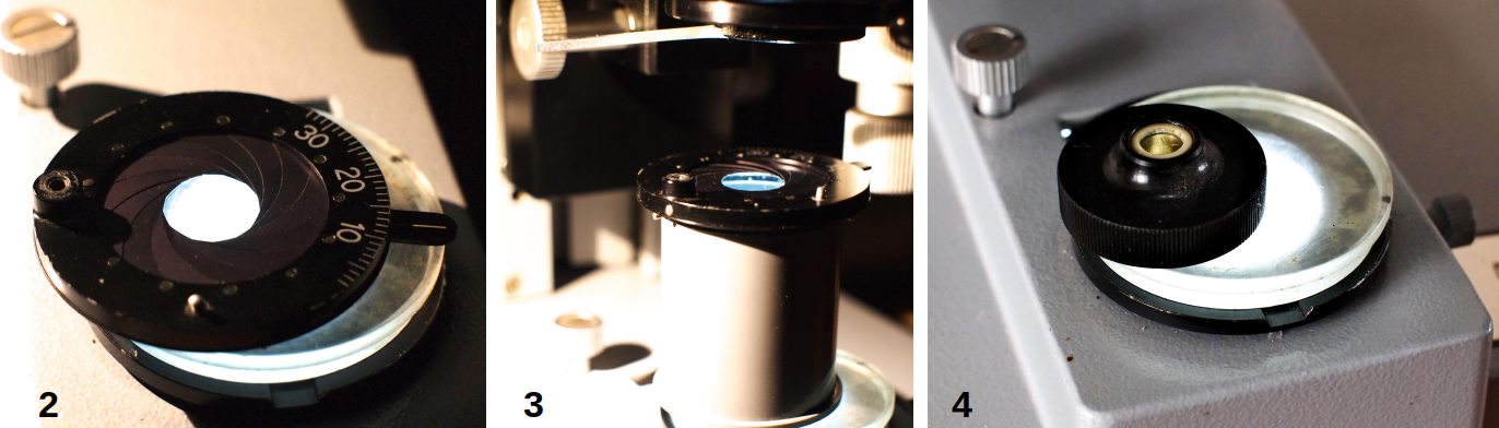
Fig.1. Methods to achieve oblique illumination. A: Darkfield stop of 18 mm in diameter in filter holder. B: Shifting the auxiliary lens. C: Shifting the filter holder. D: Tilted condenser top lens.
The following images of Pleurosigma angulatum were made using different settings. The effect of the settings (numbered 1 to 6) on the illumination at the back lens of the objective as seen through a phase-telescope is shown in figure 5.
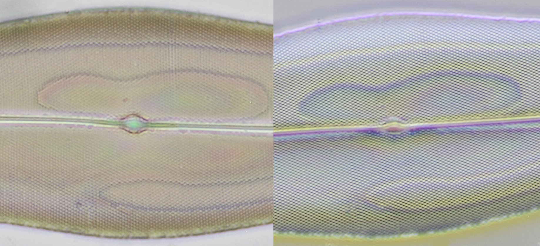
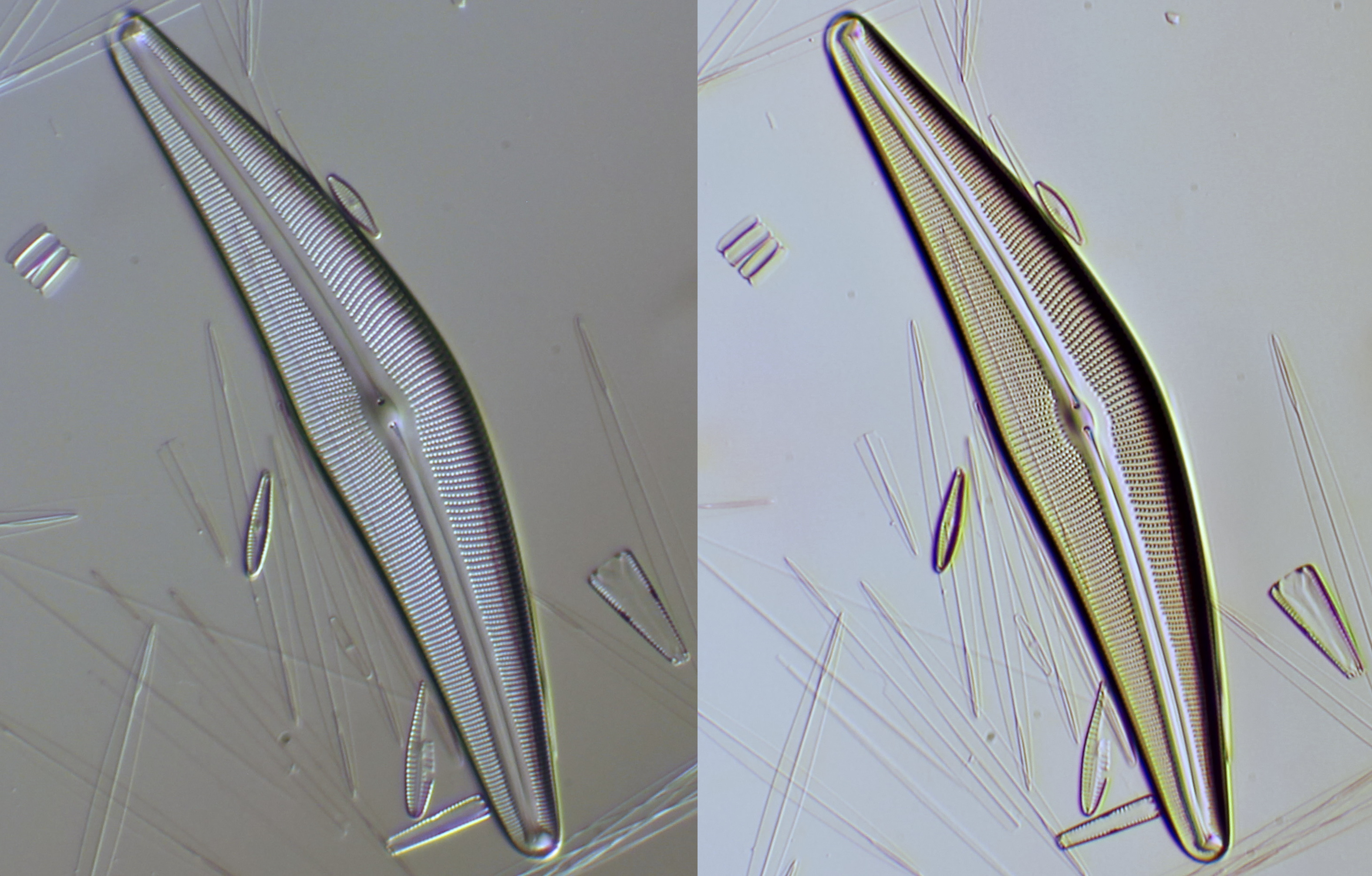
Fig.2. Pleurosigma angulatum in normal bright field illumination (left, setting 1) and oblique illumination using a darkfield stop in the filter holder (right, setting 2). In the right image, the fine structure of this diatom is much more visible.
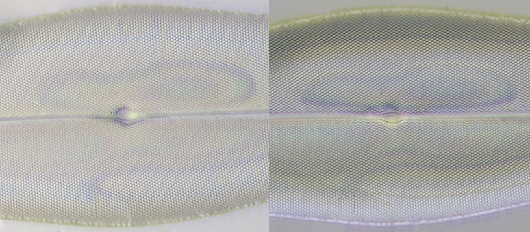
Fig.3. Oblique illumination by decentering the condenser (left, setting 3) or by shifting the auxiliary lens under the condenser (right, setting 4).

Fig.4. Oblique illumination by tilting the condenser top lens (left, setting 5) or by positioning the filter holder in the light path (right, setting 6).
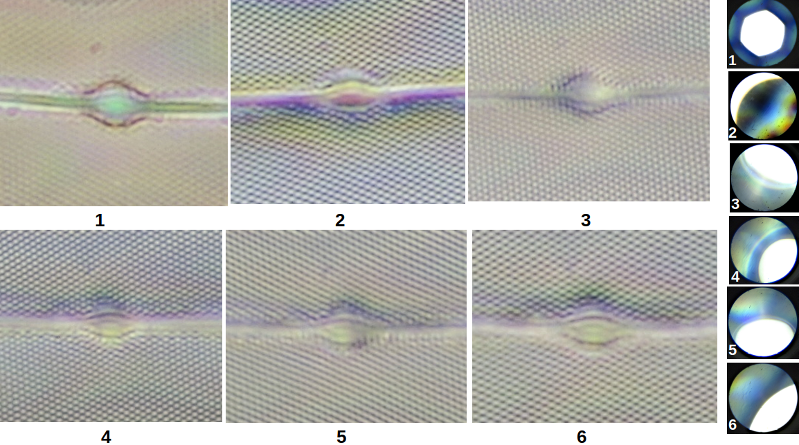
Fig. 5. Overview of the different settings. 1: Bright field illumination. 2: Darkfield stop. 3: Decentering the condenser. 4: Auxiliary lens shifted under condenser. 5: Tilted condenser top lens. 6: Filter holder positioned in light path. On the far right of the picture, the illumination at the back lens of the objective is seen, photographed using a phase telescope.
I personally find that in the experiment above the best contrast and resolving power was obtained by shifting the auxiliary lens (setting 4) or using a darkfield stop in the filter holder (setting 2). In my opinion, these two methods proved to be best in resolving the structure of Pleurosigma angulatum. On the other hand, decentering the condenser (setting 3) produced the least color artifacts and a nearly gradient-free oblique illumination was obtained.
In addition to improved contrast and resolving power, oblique illumination has a positive effect on the depth of field. At the same time, a pseudo-relief is created which can be clearly seen in the following image.
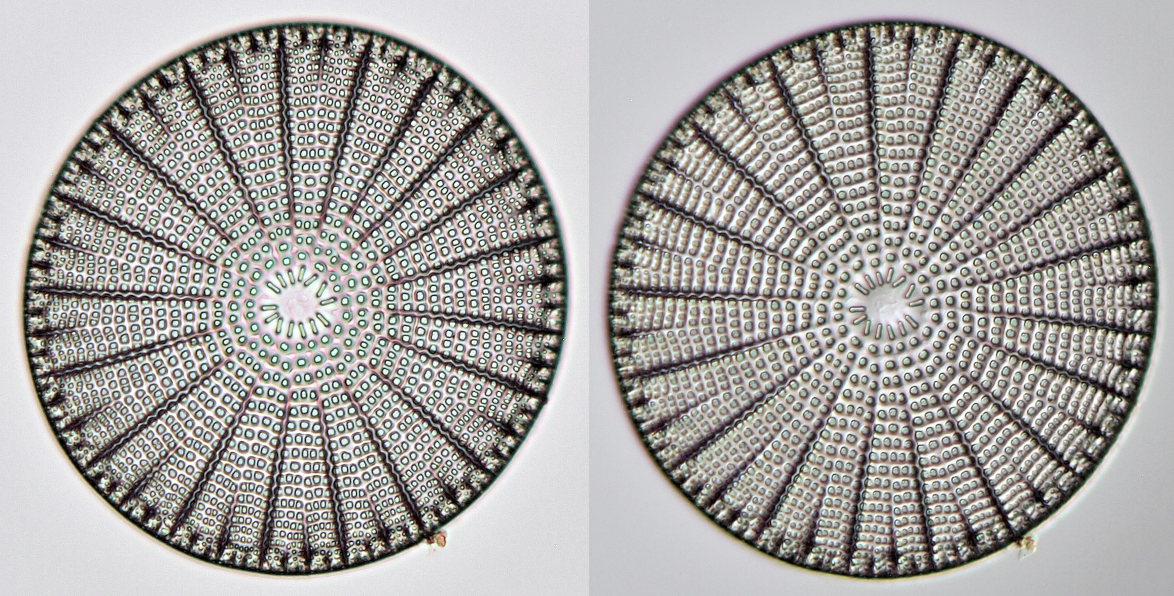
Arachnoidiscus ehrenbergii fotographed in brightfield (left) and oblique illumination (right). Objective: Zeiss-Winkel 25/0.45.
The "field lens method"
I find that the most satisfactory results are obtained by placing a lens on the light opening. The lens I used for this came from a Carl Zeiss Jena (1.2 N.A.) Abbe condenser. I unscrewed the top lens and placed the bottom lens (the field lens) asymmetrically on the light opening. Of course, field lenses from other condensers can also be used.
The following experiments were done with a Zeiss Standard GFL, in which I also compared the field lens method with a more common method for obtaining oblique illumination: shifting the aperture diaphragm. The latter is very easy when you have a phase contrast condenser: the bright field position is turned to the left or right so that the light comes from one side. In this way, a good oblique illumination can be obtained very easily.
With the field lens method, the condenser is turned all the way up and the aperture diaphragm is opened completely. The diaphragm at the light source (the field diaphragm, if present) is also opened to its maximum. A diffusing filter is placed on the light opening and then the field lens is placed and shifted until a satisfactory result is achieved. I set the position of the field lens in such a way that a not too strong illumination gradient is created in the image while at the same time enough depth of field is created. With a little experimentation results are achieved that are very similar to Differential Interference Contrast (DIC). In my opinion, the field lens method gives a more refined oblique illumination with fewer artefacts. In addition, the contrast is less high, which makes some details more visible. The image also seems to have more depth compared to the usual method. These are all subjective impressions of course. The best thing to do is to try it out yourself and observe carefully what happens using different slides.
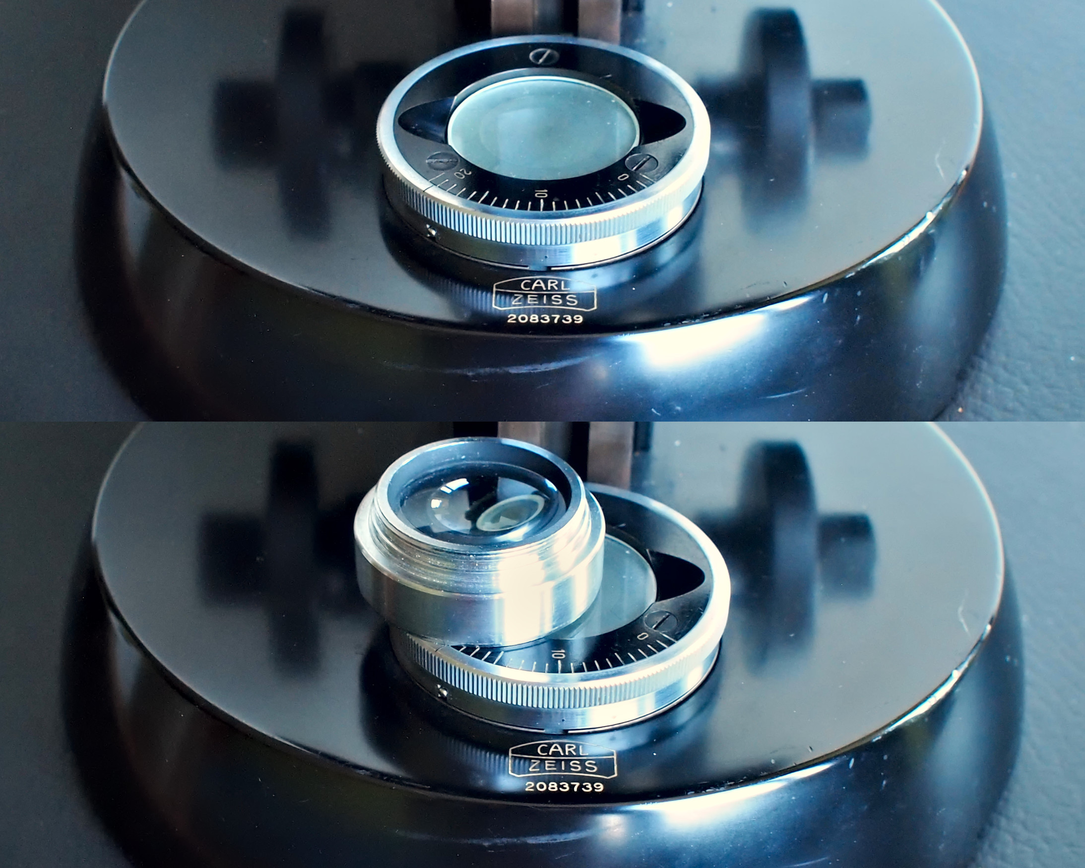
The field lens method for obtaining oblique illumination with a Carl Zeiss Standard GFL. Oblique illumination is obtained here by influencing the light rays at the light source. First, a diffusing filter is placed on the light opening (top). Then, the field lens of a condenser is placed on top of the diffusing filter and is shifted off-center to produce oblique illumination (bottom photo).
When the illumination at the back of the objective is inspected with a phase telescope, it is noticeable that the illumination with the field lens method is rather diffuse. A light gradient is created. With the usual method of obtaining oblique illumination, this looks very different: here a clearly defined part of the objective is illuminated.
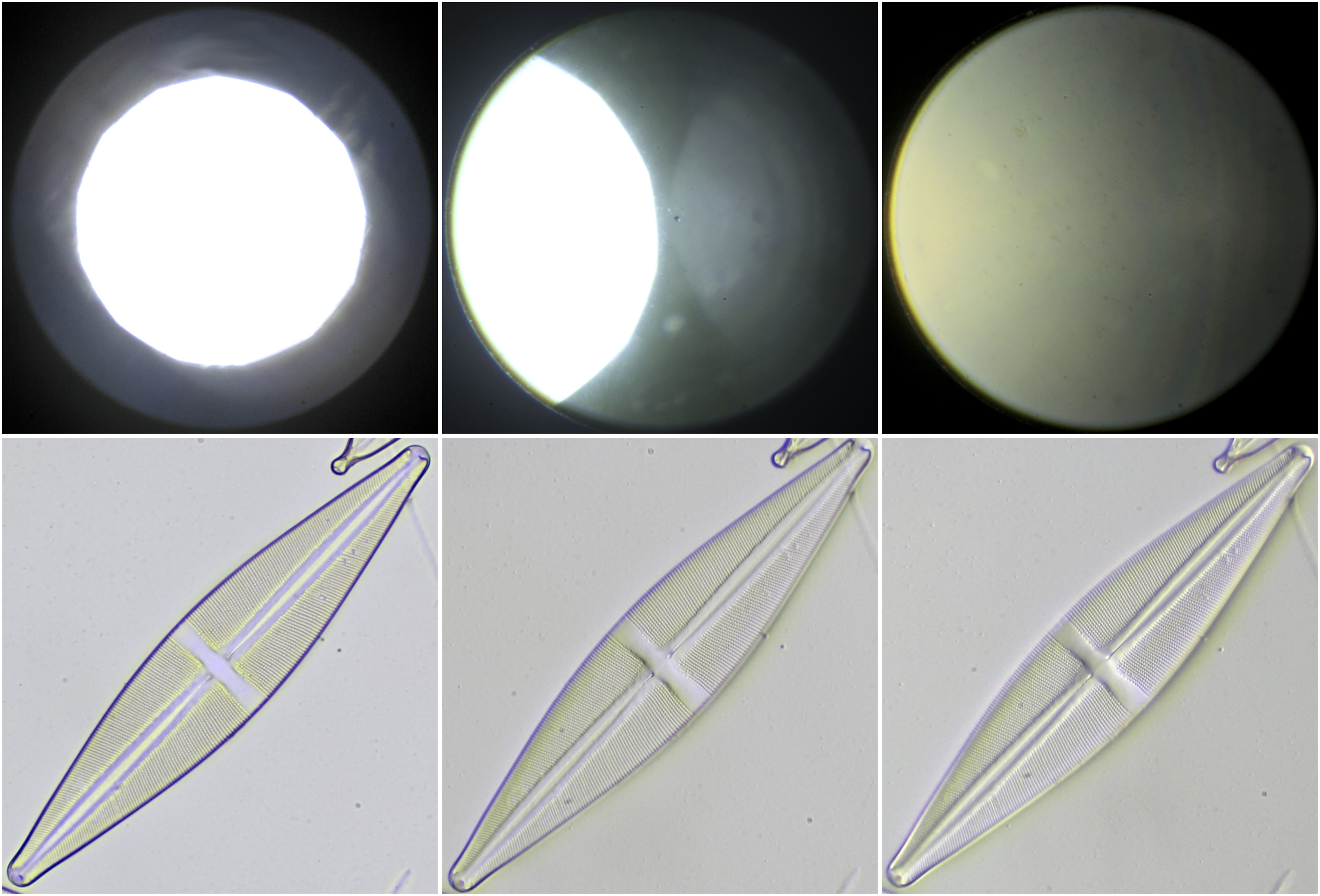
Stauroneis, photographed with Carl Zeiss 40/0.65 and different illumination methods. From left to right: normal bright field, oblique illumination by shifting the aperture diaphragm, field lens method. The upper series of photos shows the illumination of the objective as seen with a phase telescope. Note the very diffuse illumination with the field lens method. When looking at the diatom, it is noticeable that the field lens method gives less color artifacts and the object seems to have more depth here.
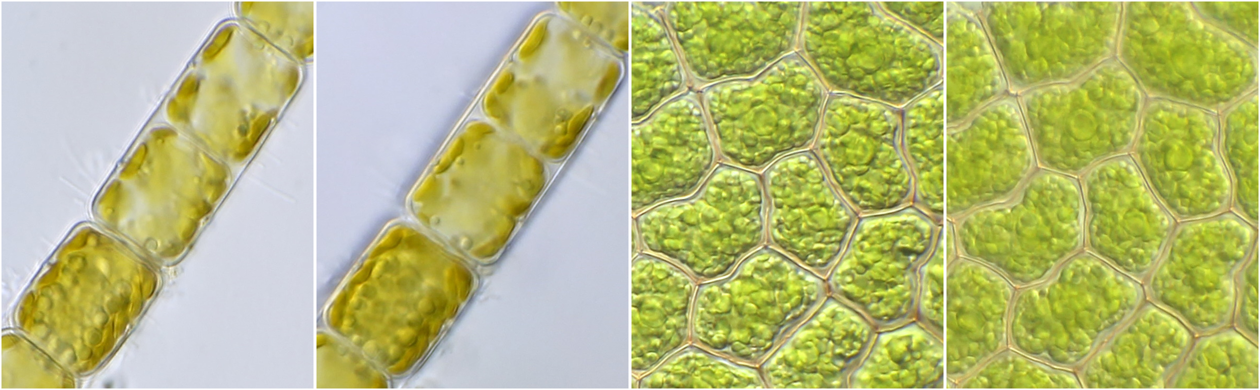
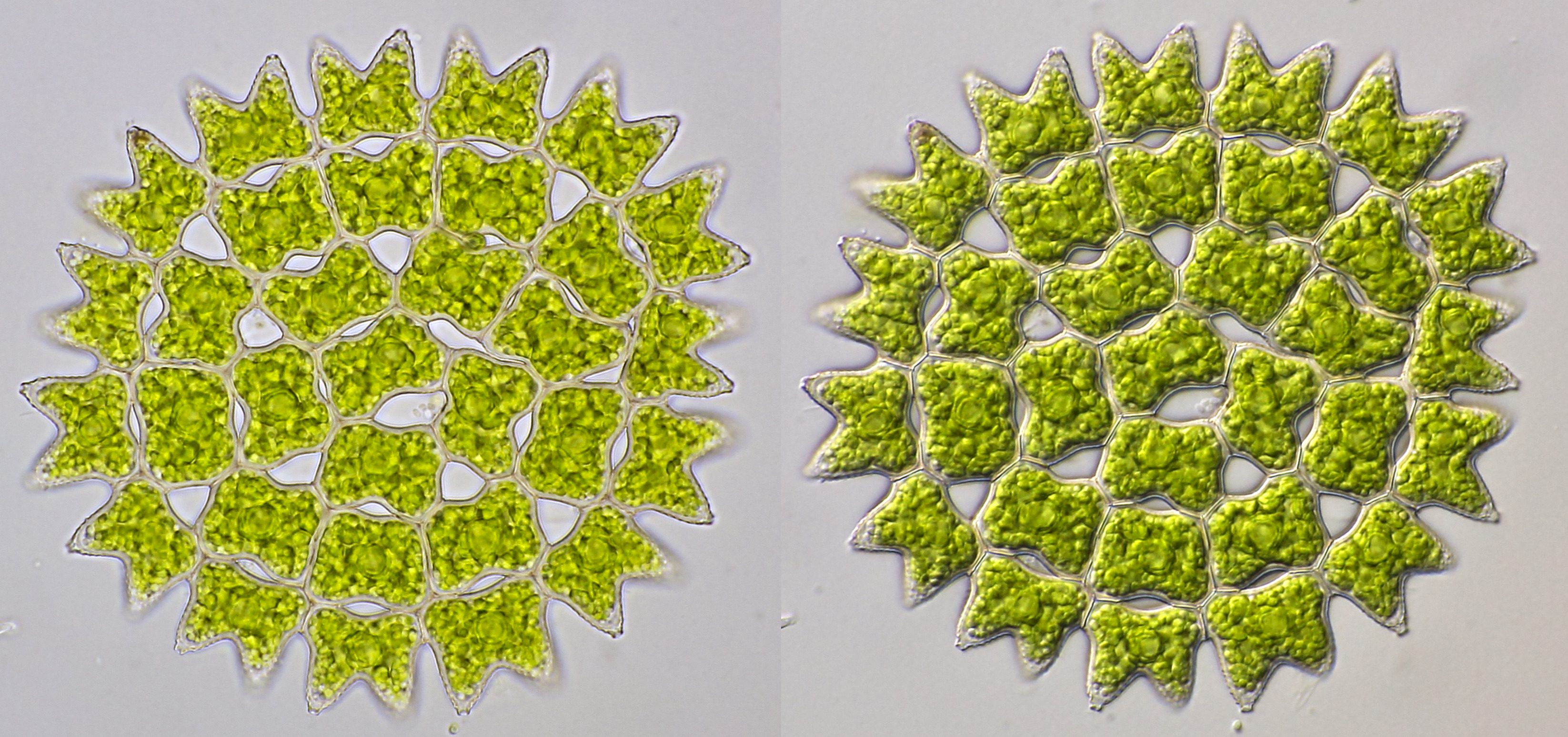
Comparison of oblique illumination by shifting the aperture diaphragm in a Carl Zeiss phase contrast condenser 0.9 (photo 1 and 3) and the field lens method (photo 2 and 4). Melosira (photo 1 and 2) and Pediastrum (photo 3 and 4) served as test objects. What is striking is that the contrast in photo 1 and 3 is higher while the depth of field is smaller there. In my opinion, the field lens method gives a more subtle oblique illumination with less harsh contrasts at the edges. The image can sometimes be a bit faint as can be seen in photo 4, but with some image editing the contrast can be adjusted to your liking. Objective: Carl Zeiss Apo 40/1.0.
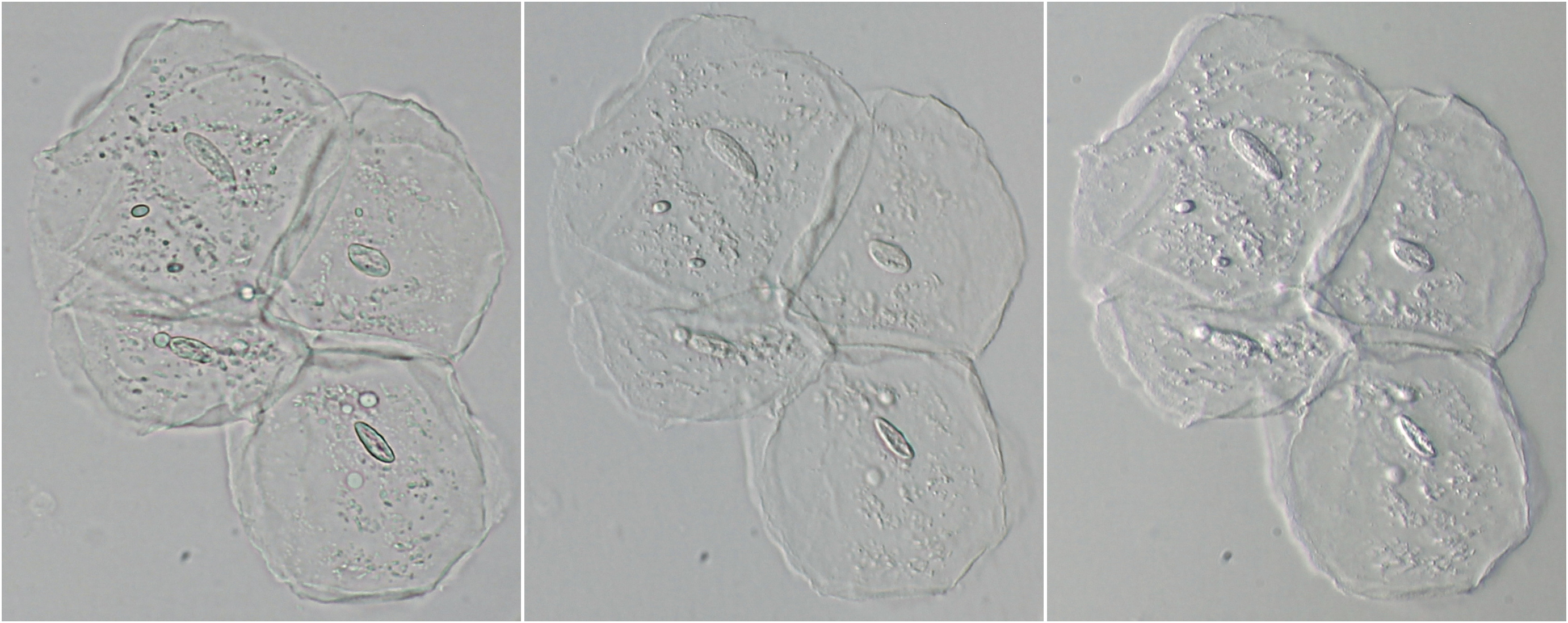
Cheek epithelial cells photographed in bright field (left), oblique illumination by shifting the aperture diaphragm (middle) and oblique illumination by field-lens method (right). The images were taken with a Leitz Laborlux-12 with phase contrast condenser 1.2 and NPL Fluotar 25/0.55.
Cymbella photographed with Carl Zeiss Plan 25/0.45. Left: oblique illumination using the field lens method. Right: oblique illumination by shifting the aperture in the phase contrast condenser. The difference in effect is significant here.
Pediastrum, photographed in bright field (left) and oblique illumination (right, single image) as described above. Objective: Carl Zeiss Apo 40/1.0. For the image with oblique illumination, the aperture of the objective was reduced to approximately 0.8 by means of the iris in the objective. Camera: Canon EOS 600D.
Leitz Dialux II
I have experimented a bit with oblique illumination on the Leitz Dialux II, using the Leitz Fluotar 25/0.55 objective. The diatom Cymbella was used as a test-object and the oblique illumination was realised in 5 different ways:
1: The brightfield position of fasecontrast condensor 402a was decentered.
2: A diffuser was placed on the lamp-opening and a diaphragm was, slightly decentered, placed on top of it.
3: As in method 2, but now the diaphragm was raised by putting it on a plastic tube.
4: A diffuser was placed on the lamp-opening and on top of this a black disc was placed in a decentered way. The black disc was a course focussing knob of a microscope.
5: Decentering the darkfield stop of fasecontrast condensor 402a.
Introducing oblique illumination on the Leitz Dialux II with methods 2, 3 en 4.

Experimenting with oblique illumination on a slide of Cymbella. A: normal brightfield illumination. 1-5: oblique illumination with the different methods described above.
Conclusion
Oblique illumination enhances contrast and improves the visibility of fine details and can be achieved with simple means. There are many different ways to create oblique illumination and each method has it’s own characteristics. The depth of field can be increased considerably with oblique illumination. It gives a more spatial impression of the object than normal brightfield illumination and the visibility of some details will be enhanced. If you look at photos taken with oblique illumination, there may be a sudden reversal of the relief; cavities suddenly look like bulges. This is an optical illusion. It is good practice to always take a picture in brightfield and compare it with the image taken with oblique lighting.