
Introduction
Imagine a landscape with hills, valleys, and dales, covered exclusively with mosses. That is what it must have looked like a long time ago. It is believed that mosses (Bryophyta) were the first land plants on Earth. Unlike ‘true’ land plants, mosses don’t have roots. Mosses have root-like structures called rhizoids with which they attach themselves to a substrate. The primary function of rhizoids is therefore not the absorption of water and nutrients; in this respect, they differ from true roots. Water and nutrients are absorbed by the entire plant via diffusion, and with the numerous small leaves, this covers a large surface area. A moss can survive for a very long time in a dried-out state, and as soon as water becomes available again, the plant revives.
Moss propagation takes place via spores, and its life cycle consists of a so-called gametophyte and sporophyte. The gametophyte is the haploid phase of the plant, while spores are formed during the diploid phase within the sporophyte. From a germinating spore arises the protonema, which is the precursor from which the haploid plant is formed.
Mosses are rewarding subjects for microscopy. A single small leaf turns out to be a miniature structure under the microscope. Moss leaves are almost always only one cell layer thick; in some species, the leaf margin can be two or more cell layers thick. The very small cells are often packed with chloroplasts, and you sometimes wonder how there can be room for anything else. During a walk through the forest, I usually carry a few small jars with me so that I can collect interesting mosses along the way to examine later under the microscope.
Identifying mosses is a specialized skill, and it is often difficult to name the species. Consequently, the name of the moss is frequently missing from the images on this page.
Mosses around the house
Mosses grow wherever it is damp and there is enough light. You only have to walk outside, and even if you have a tiled garden, there is a good chance that moss is growing on those tiles. I found the following mosses in various backyards and balconies.
Leaves of moss growing on a paving stone. Photographed with a Zeiss-Winkel horseshoe stand and Winkel-Zeiss 13/0.32 objective. Left: brightfield image. Right: oblique illumination.
Moss leaves from a moss in the same backyard as above, also growing on a paving stone. Brightfield images with Zeiss-Winkel 10/0.25 (left) and Zeiss-Winkel 40/0.65 (right).
A moss from our garden in Zevenaar photographed with Zeiss 25/0.45 and various illumination methods. From left to right: dark field, oblique lighting, and polarised light.
Moss leaf of the same moss as above, from the garden in Zevenaar. Photographed with oblique lighting with a Zeiss 25/0.45 objective.

A moss growing in a flowerpot on the balcony in Amsterdam. Left: macro image. Middle and right: photomicrographs taken with a Carl Zeiss Planachro 100/1.25. The chloroplasts are clearly visible here, and particularly in the image on the far left, starch granules are visible within the chloroplasts.

Another moss from the balcony in Amsterdam. Left: macro. Center and right: images taken using circular oblique lighting with an Olympus Plan 20/0.40. This illumination method creates some depth. Dividing chloroplasts are clearly visible.
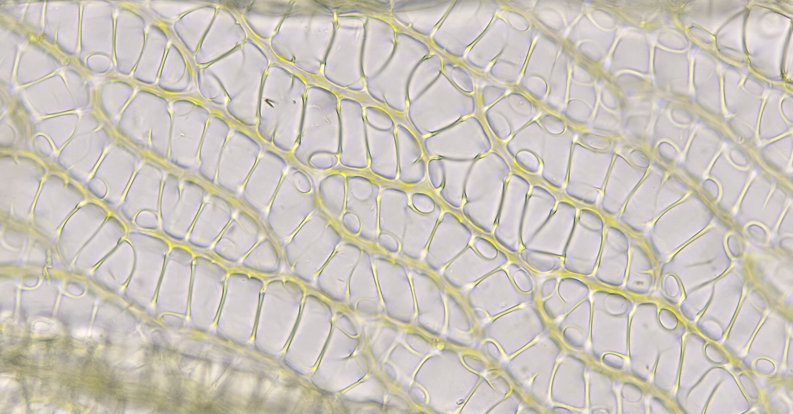
Cells in the leaf of a moss in our greenhouse at the garden park. The image was taken using darkfield illumination with a Leitz Planapo 25/0.65. It appears as if you are looking deep into the cells due to the spatial effect created by darkfield.
Tortula
Het geslacht Tortula omvat de kronkeltandmossen. Deze mossen bevinden zich vaak op muren en boomstammen. De eerste 5 afbeeldingen hieronder zijn afkomstig van een mos dat ik aantrof op een muur aan de Maaskade in Kessel. De laatste afbeelding is gemaakt van een mos uit mijn tuin in Zevenaar.
Tortula, macroscopisch beeld links en microscopisch beeld met objectief 4/0.10 rechts.
Tortula, gefotografeerd met Zeiss-Winkel 10/0.25.
De haren van Tortula zijn hier duidelijk zichtbaar. Objectief: Zeiss-Winkel 10/0.25.
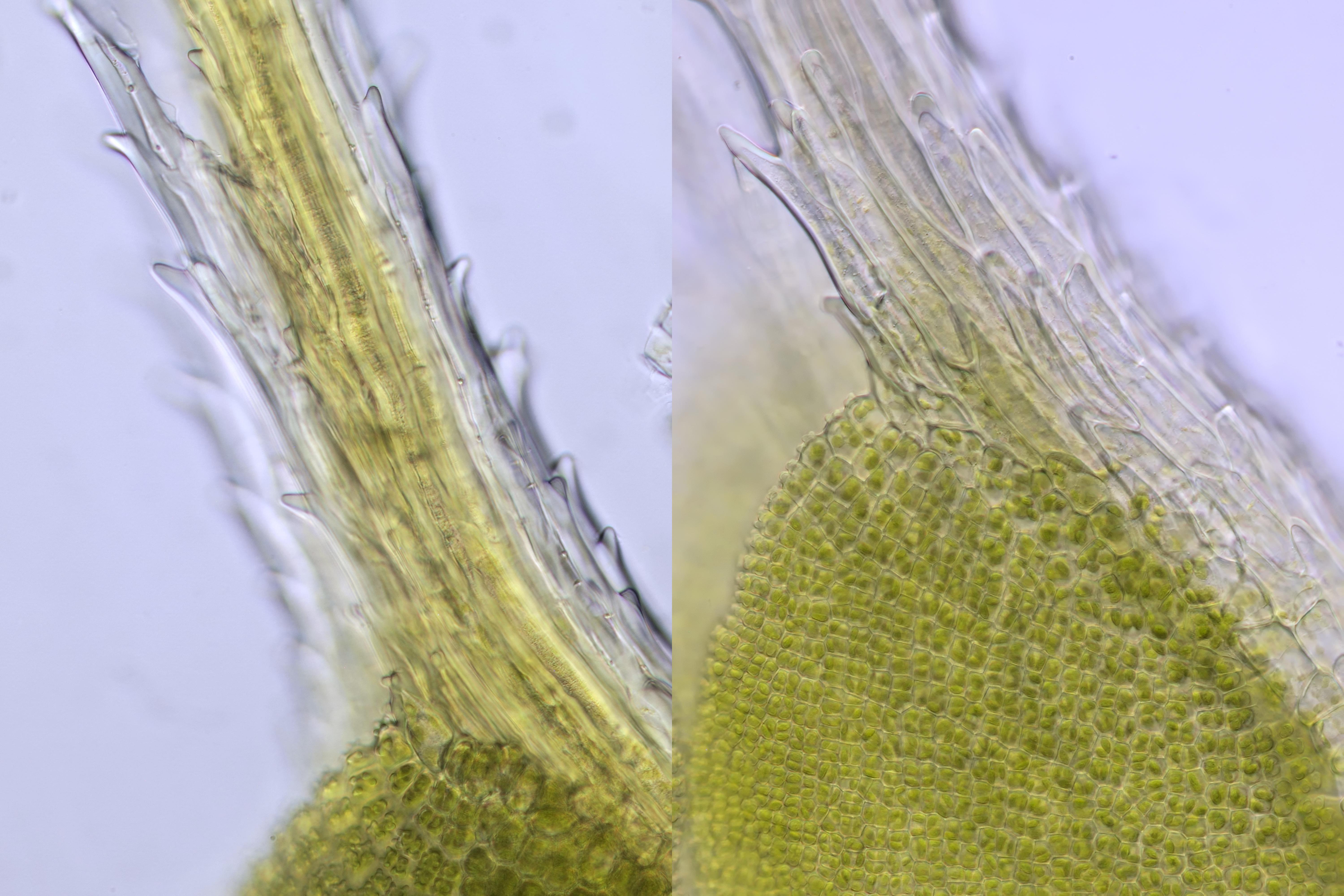
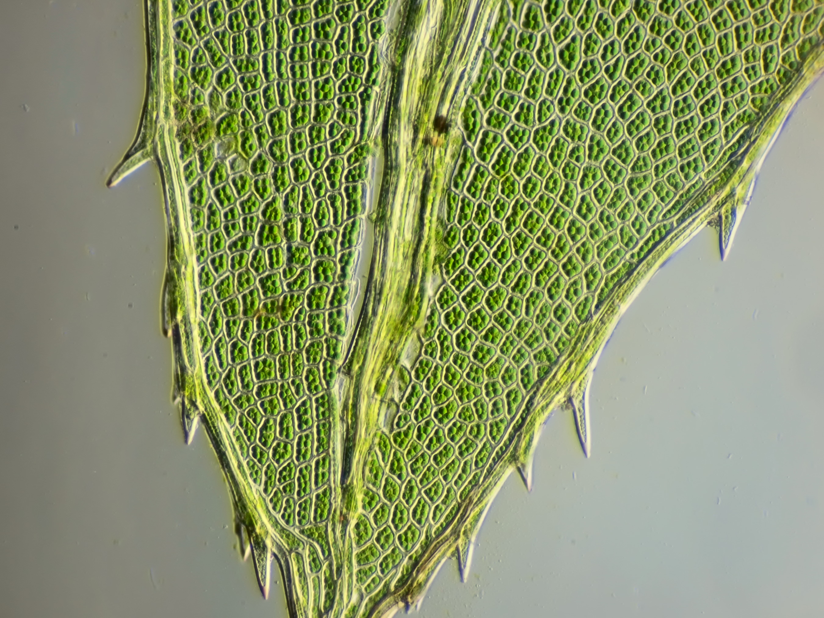
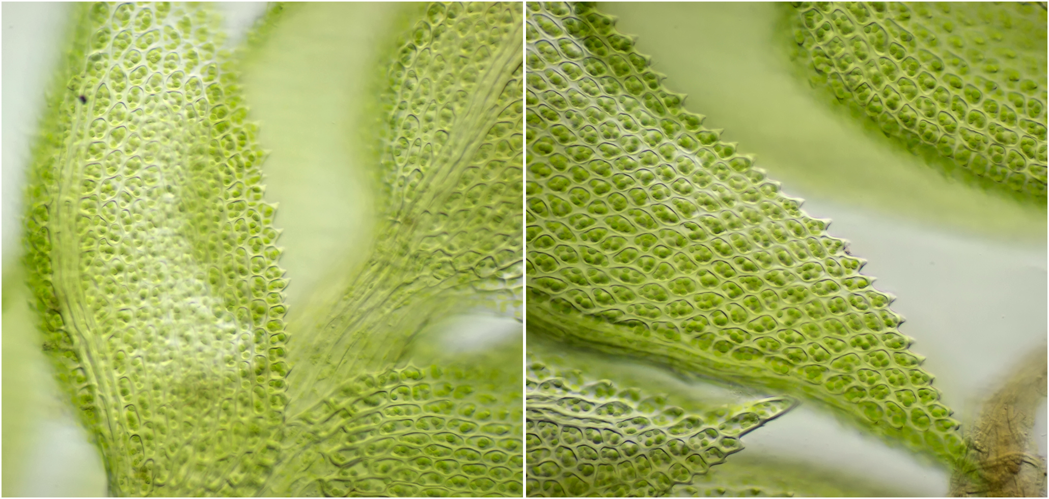
Bladuiteindes van Tortula gefotografeerd met Zeiss-Winkel 40/0.65.
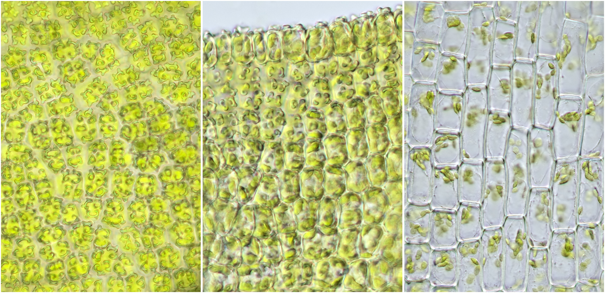
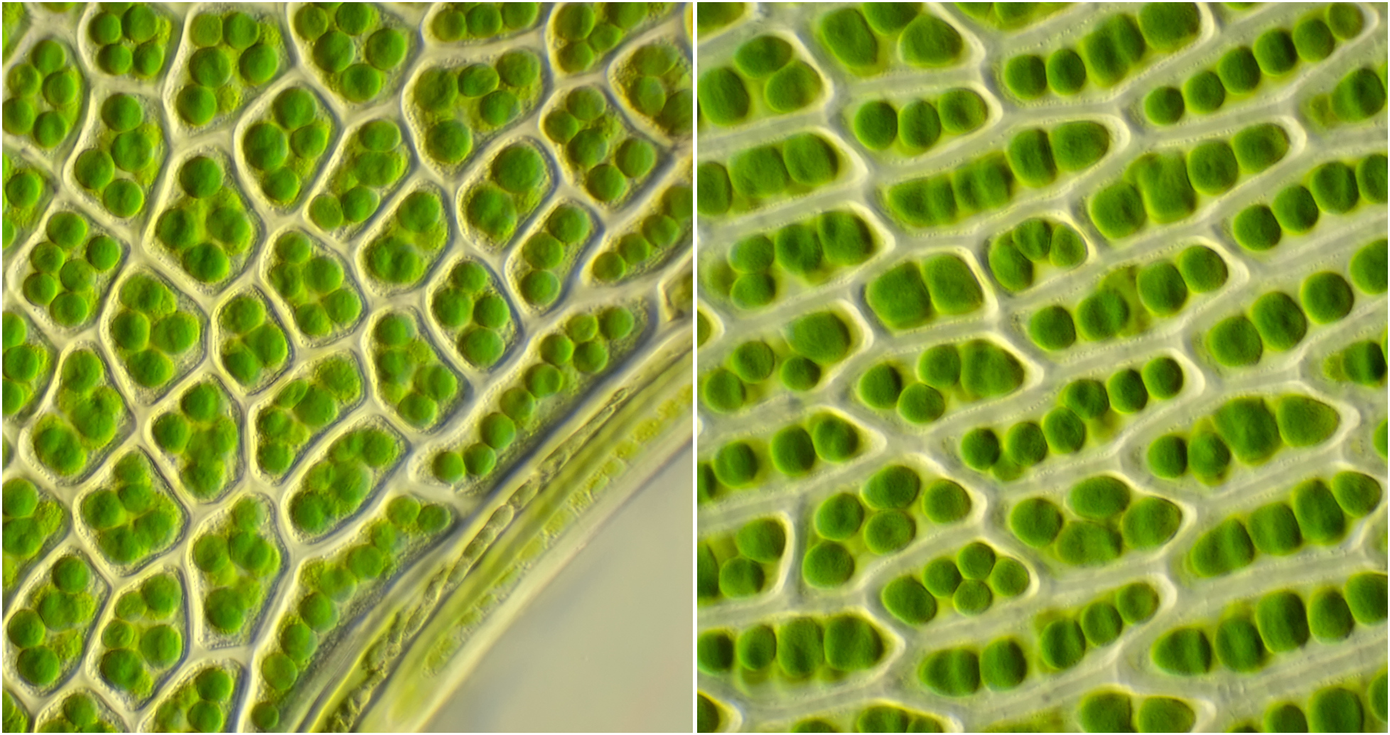
Links en midden: structuur van het bladoppervlak van Tortula waarbij de zogenaamde papillen duidelijk zichtbaar zijn. Geheel rechts: onderaan het blad is vorm en grootte van cellen anders dan bovenaan in het blad. Objectief: Zeiss-Winkel 40/0.65.
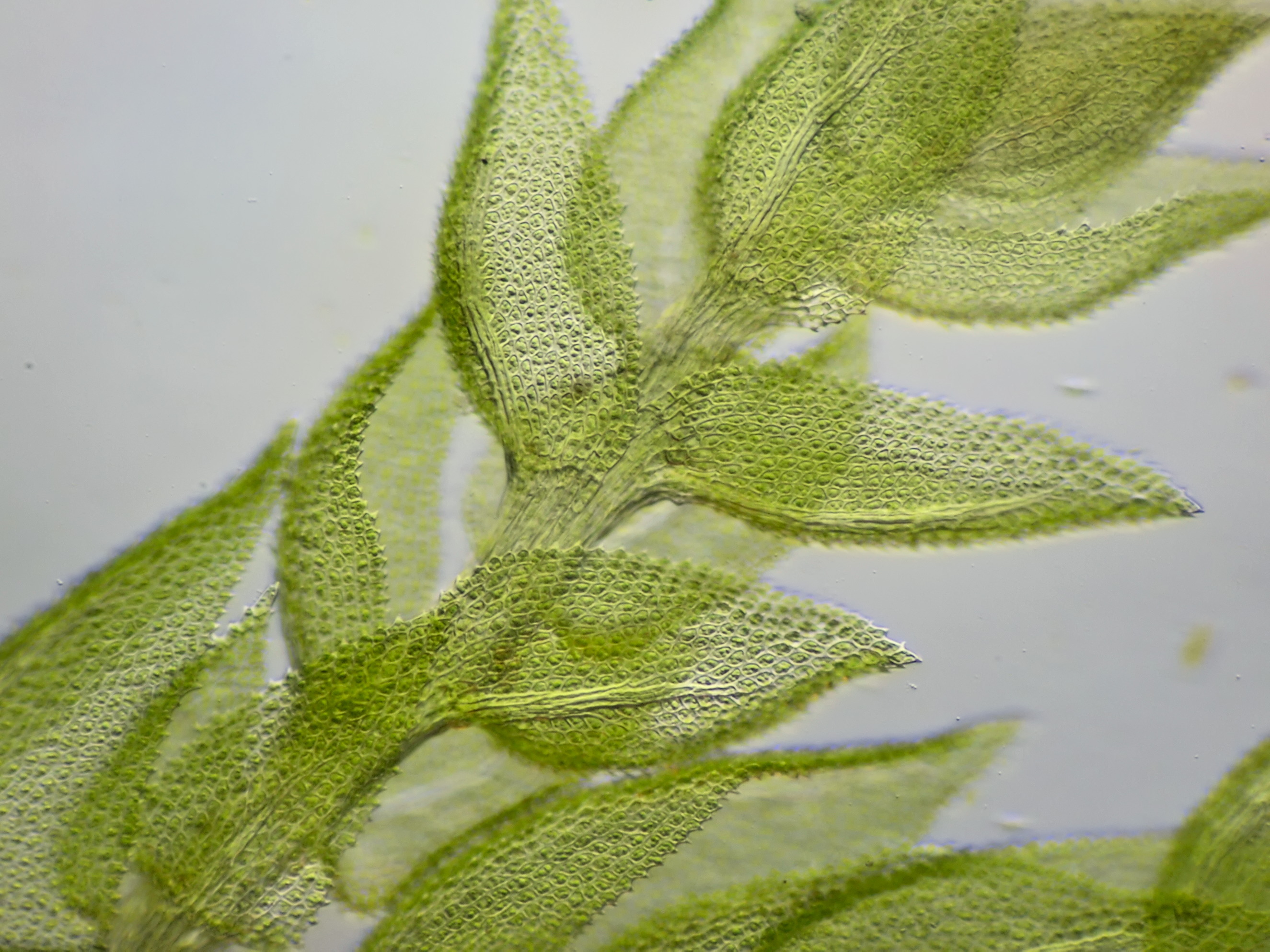
Tortula uit onze tuin in Zevenaar. Dit mos groeide op het dak en was naar beneden gevallen. Links: macro-opname met Zeiss-Winkel objectief 2.5/0.06. Rechts: de structuur van het blad-oppervlak is duidelijk te zien in deze opname die gemaakt werd met Zeiss Achroplan 40/0.65.
Marchantiophyta (levermossen)
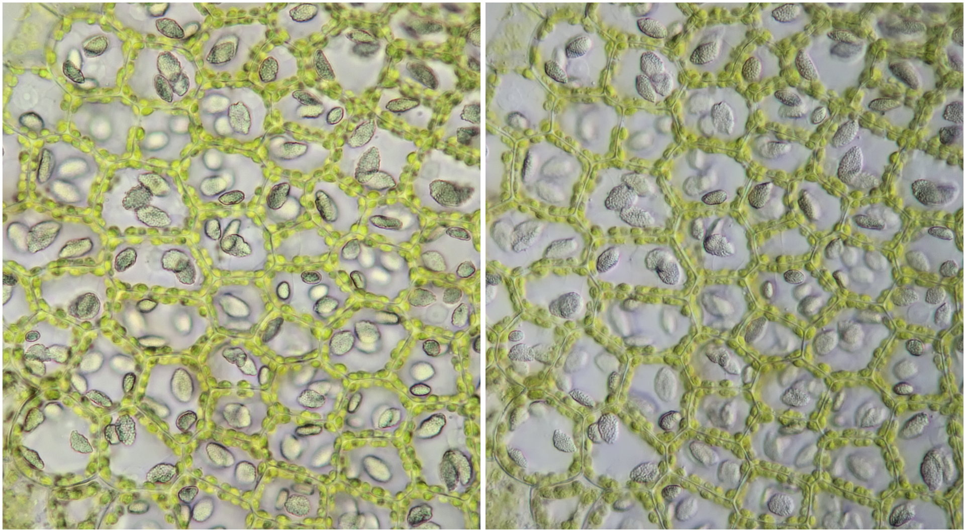
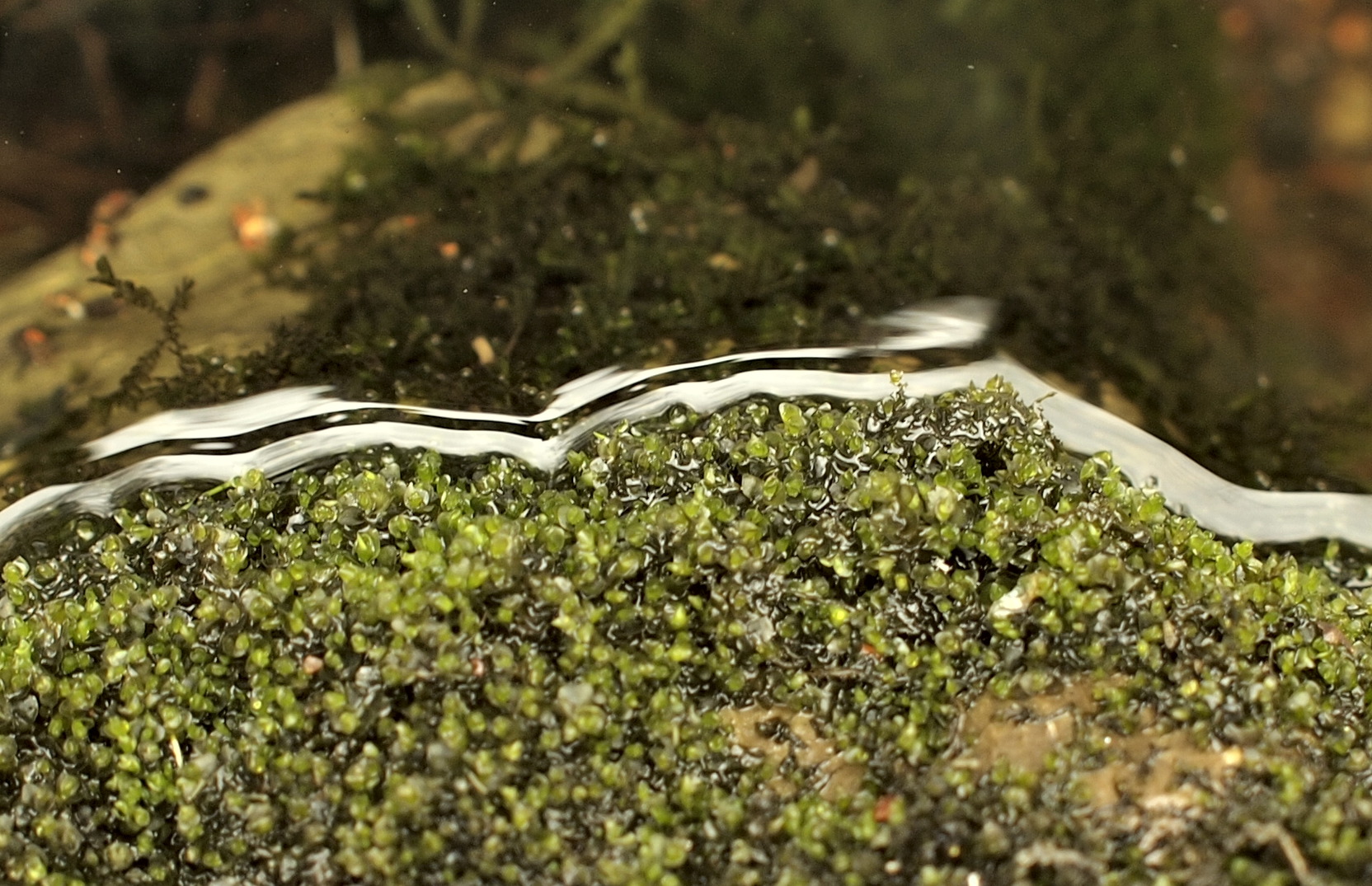
Een aparte groep van mossen wordt gevormd door de levermossen (Marchantiophyta). Toen ik diverse mossen die ik in sloten aantrof microscopisch onderzocht viel me op dat zich in de cellen korrelvormige lichaampjes bevonden. Dit bleken olie-lichaampjes te zijn en ze komen uitsluitend bij levermossen voor. Bij levermossen denkt men misschien al snel aan het typische geslacht Marchantia waaronder het parapluutjesmos valt. De mossen die onderwater voorkomen lijken echter vaak meer op gewone mossen dan op Marchantia.
Een levermos dat ik aantrof in één van de sloten in Giethoorn. Links: macro-opname van het mos. Rechts: micro-foto opgenomen met Zeiss-Winkel objectief 40/0.65. Hier zijn de grijze olie-lichaampjes in de cellen duidelijk zichtbaar.
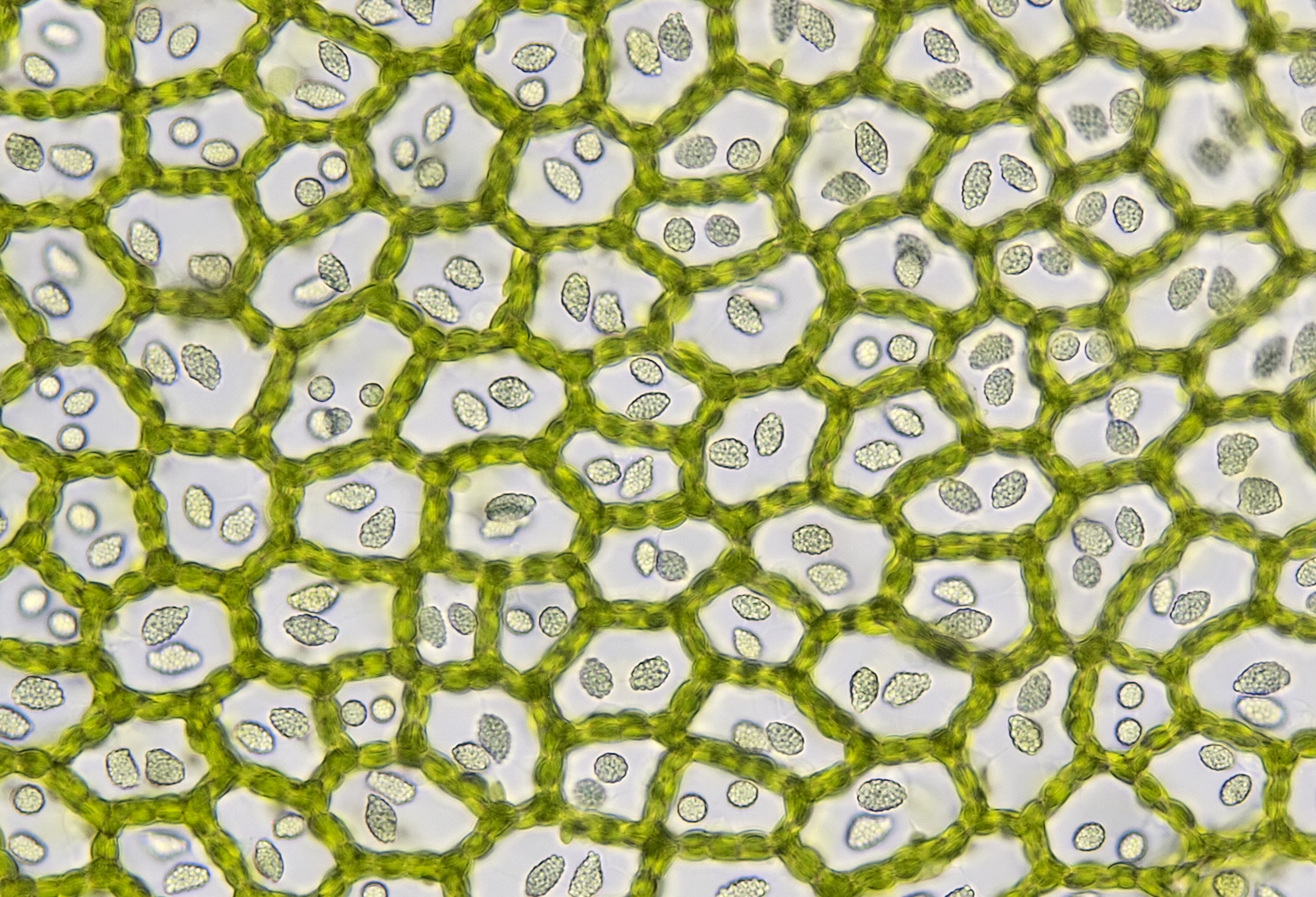
het levermos uit Giethoorn gefotografeerd met Zeiss-Winkel 40/0.65 in helderveld (links) en schuine belichting (rechts).
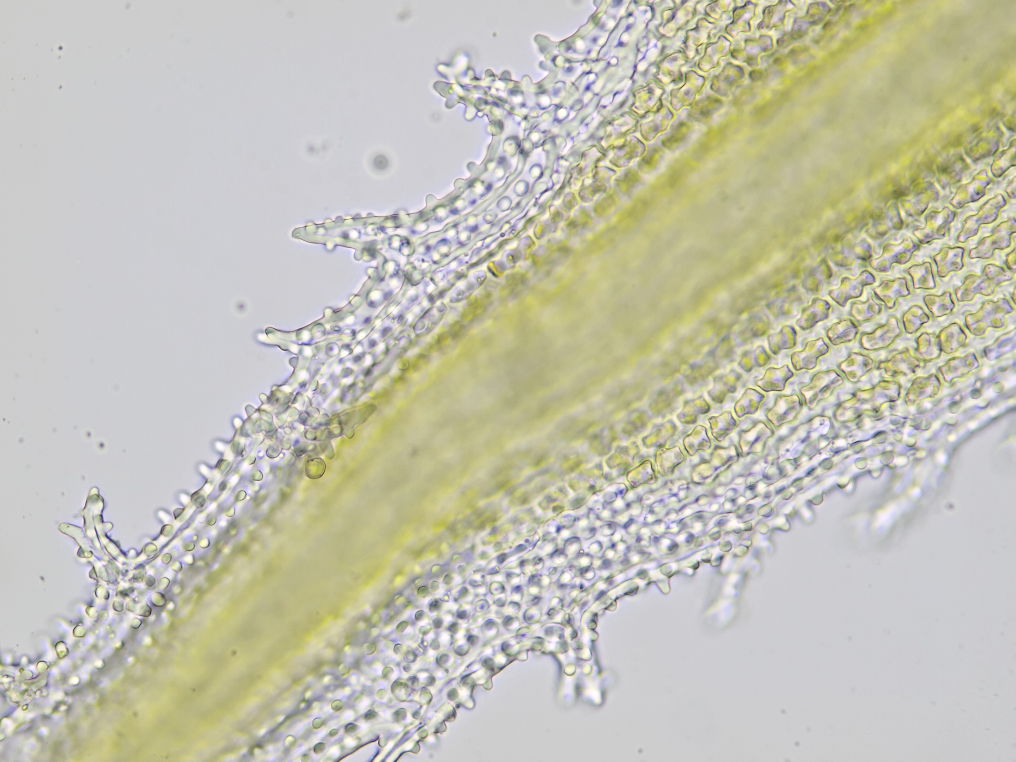
Een levermos dat ik aantrof aan de waterkant van de Rur in Heimbach tijdens een vakantie.
Micro-opname van het levermos uit de Rur in Heimbach. Cellen met wandstandige chloroplasten en olie-lichaampjes, gefotografeerd met Olympus 40/0.65 en de HSA microscoop die ik tijdens de reis had meegenomen.
Racomitrium
Racomitrium is een geslacht dat de bisschopsmutsen omvat. Een mos uit dit geslacht kwam ik tijdens een vakantie in Saarburg tegen. Hier heb ik met de Olympus HSA microscoop die ik had meegenomen foto' s van gemaakt. Wat de kenmerken betreft gaat het hier waarschijnlijk om de soort Racomitrium lanuginosum. Tot de kenmerken behoren o.a. de aanwezigheid van papillen in de de vorm van kleine bolletjes.
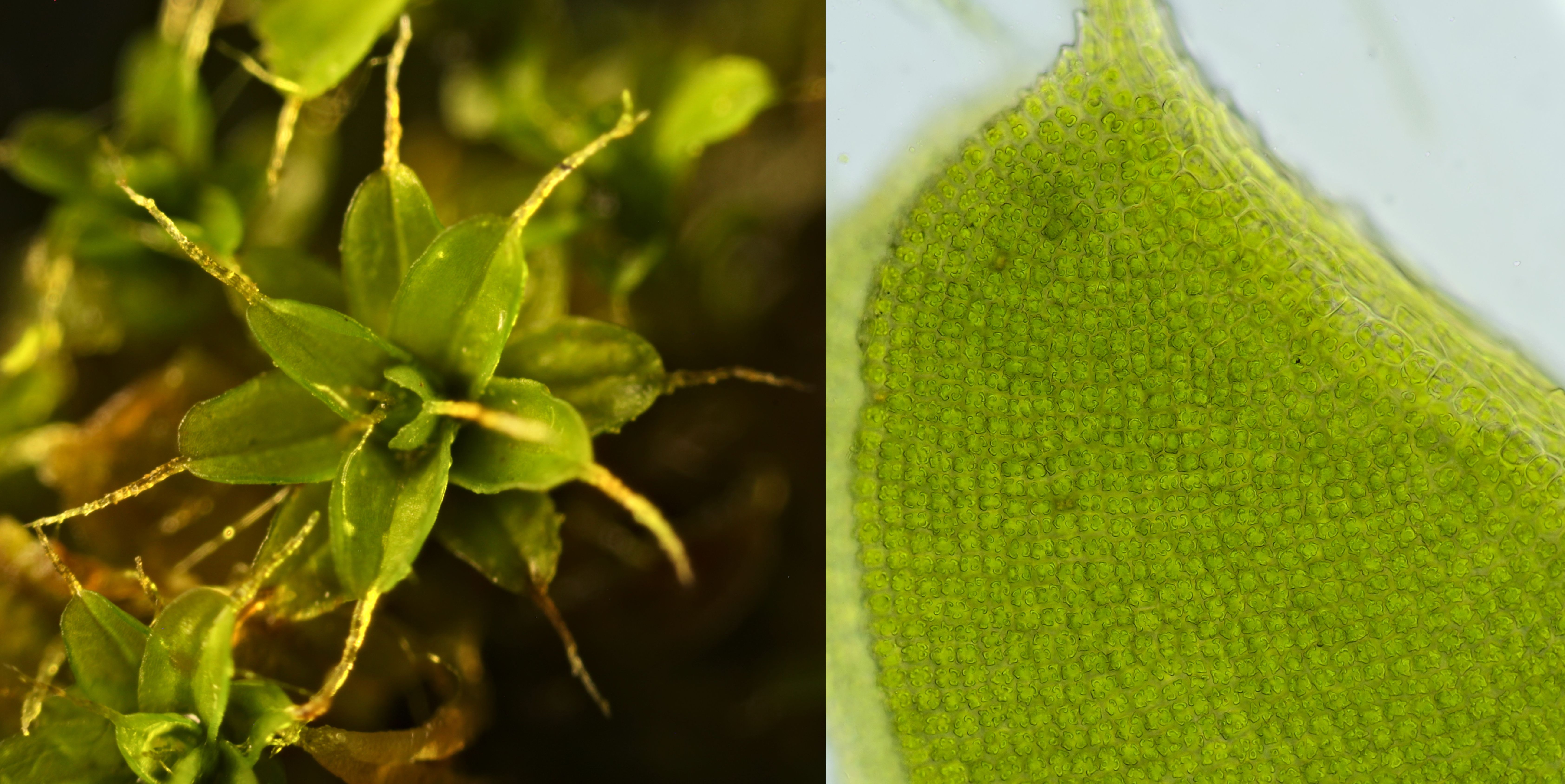
Blad-uiteinde van Rhacomitrium, gefotografeerd met Olympus objectief 40/0.65. De papillen in de vorm van kleine bolletjes zijn duidelijk zichtbaar.
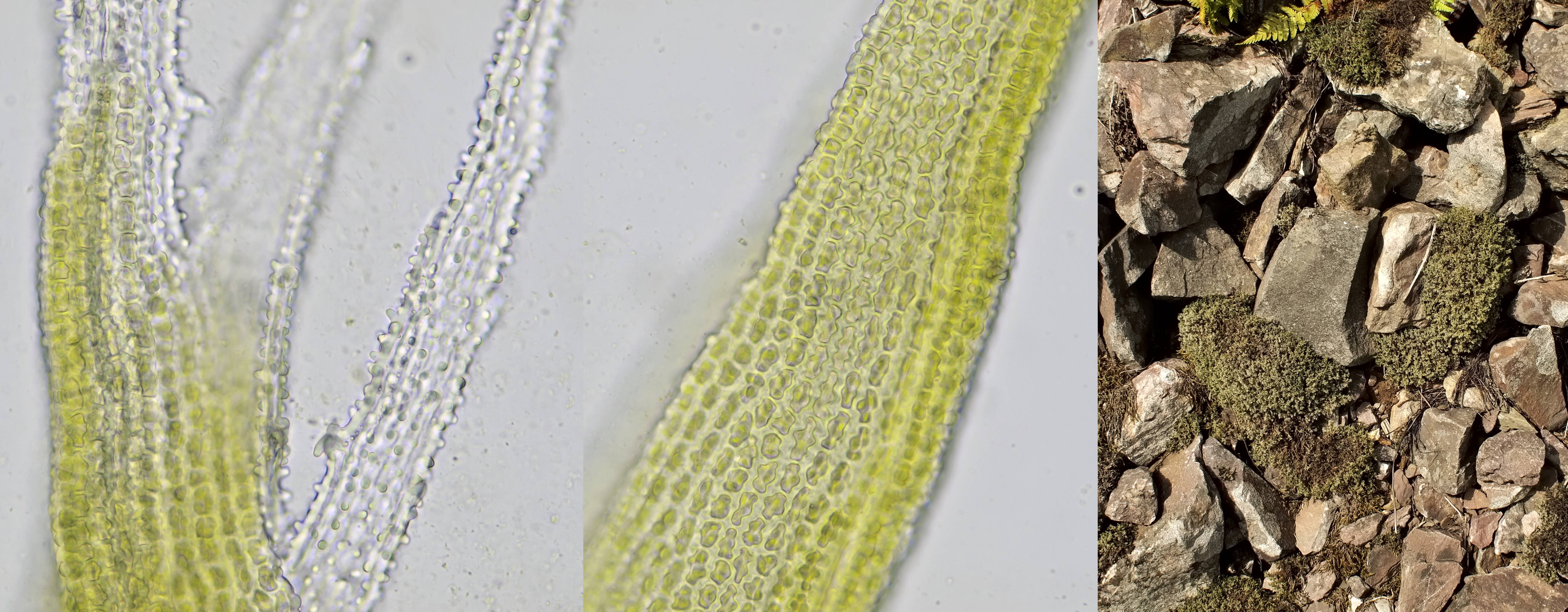
Rhacomitrium, gefotografeerd met Olympus objectief 40/0.65 (links en midden). Rechts: de vindplaats.
Dicranum
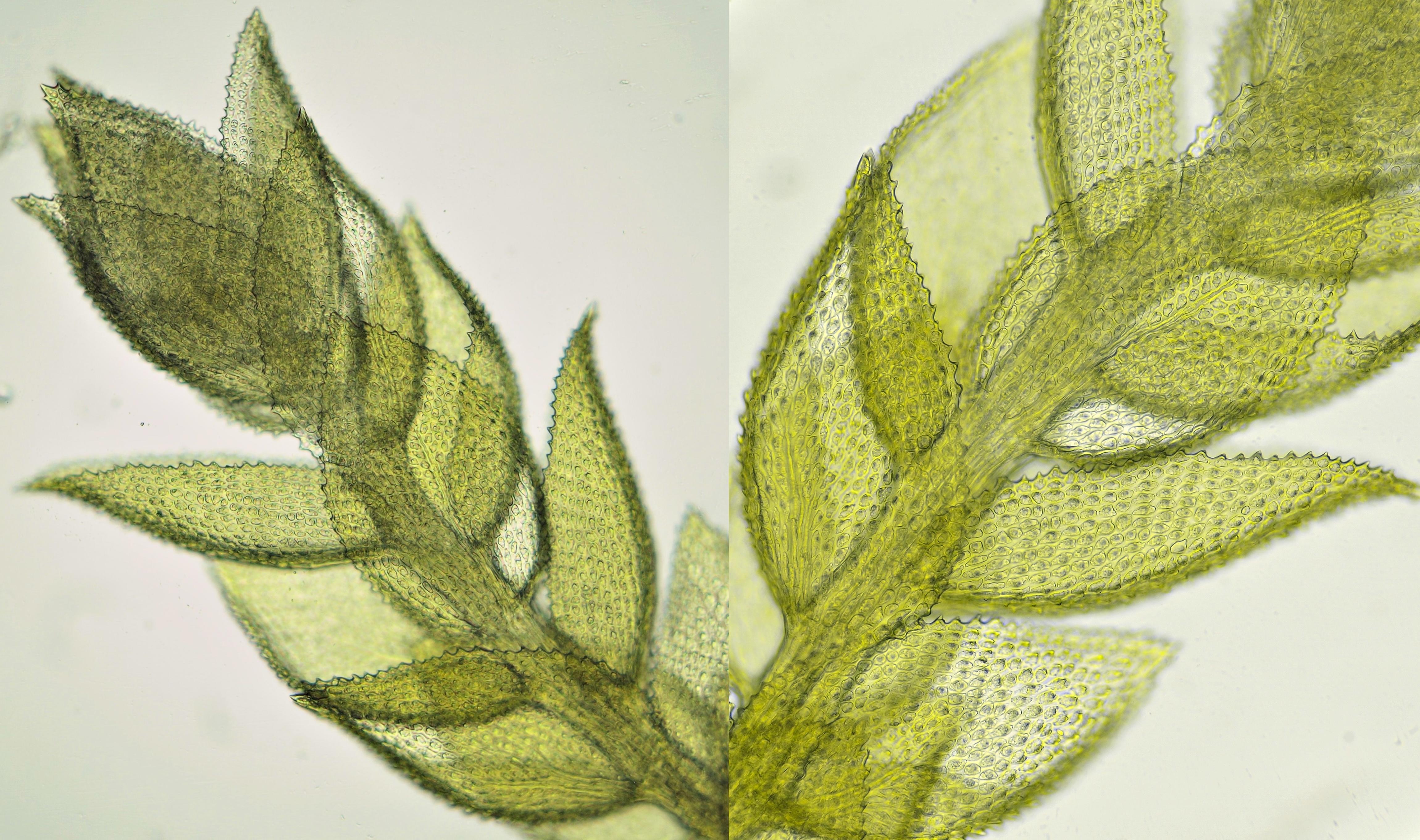
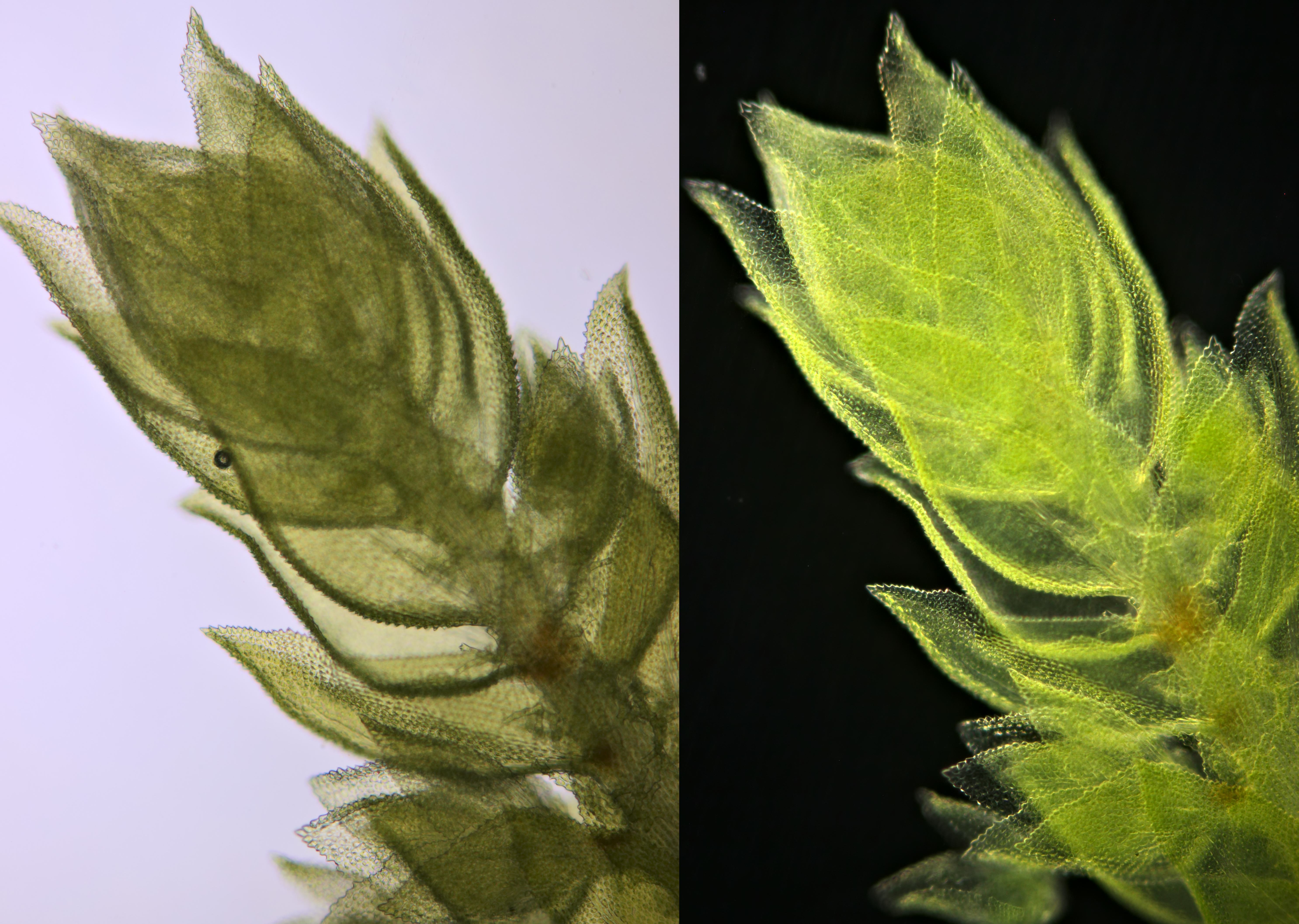
Het geslacht Dicranum omvat de gaffeltandmossen. Tijdens verschillende vakanties en wandeltochten kwam ik mossen tegen die waarschijnlijk tot dit geslacht behoorde.
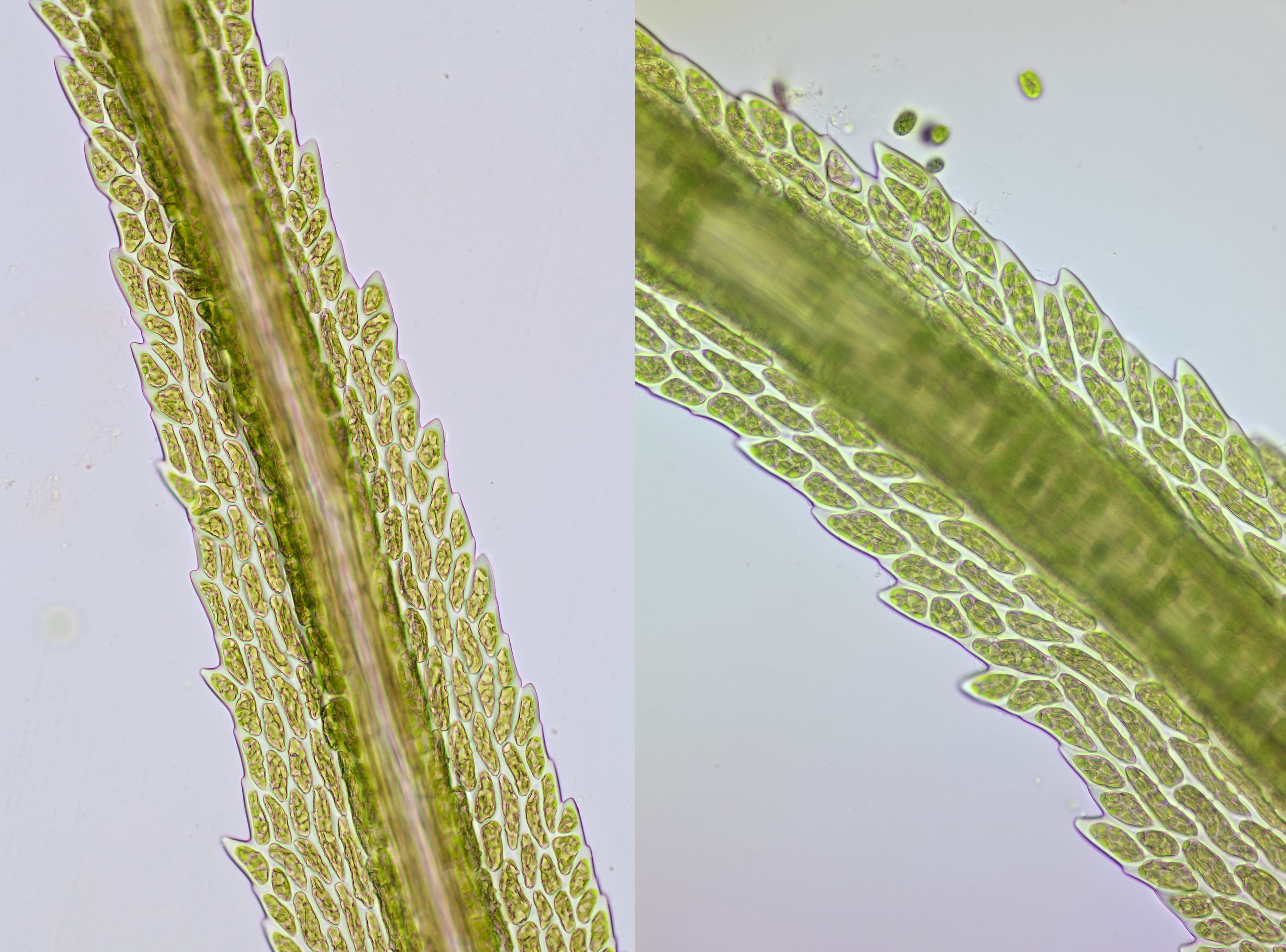
Dicranum, gevonden tijdens een vakantie in Daun (Duitsland). Gefotografeerd met Leitz EF 25/0.50 (links) en Motic-EF-N-Plan40x.
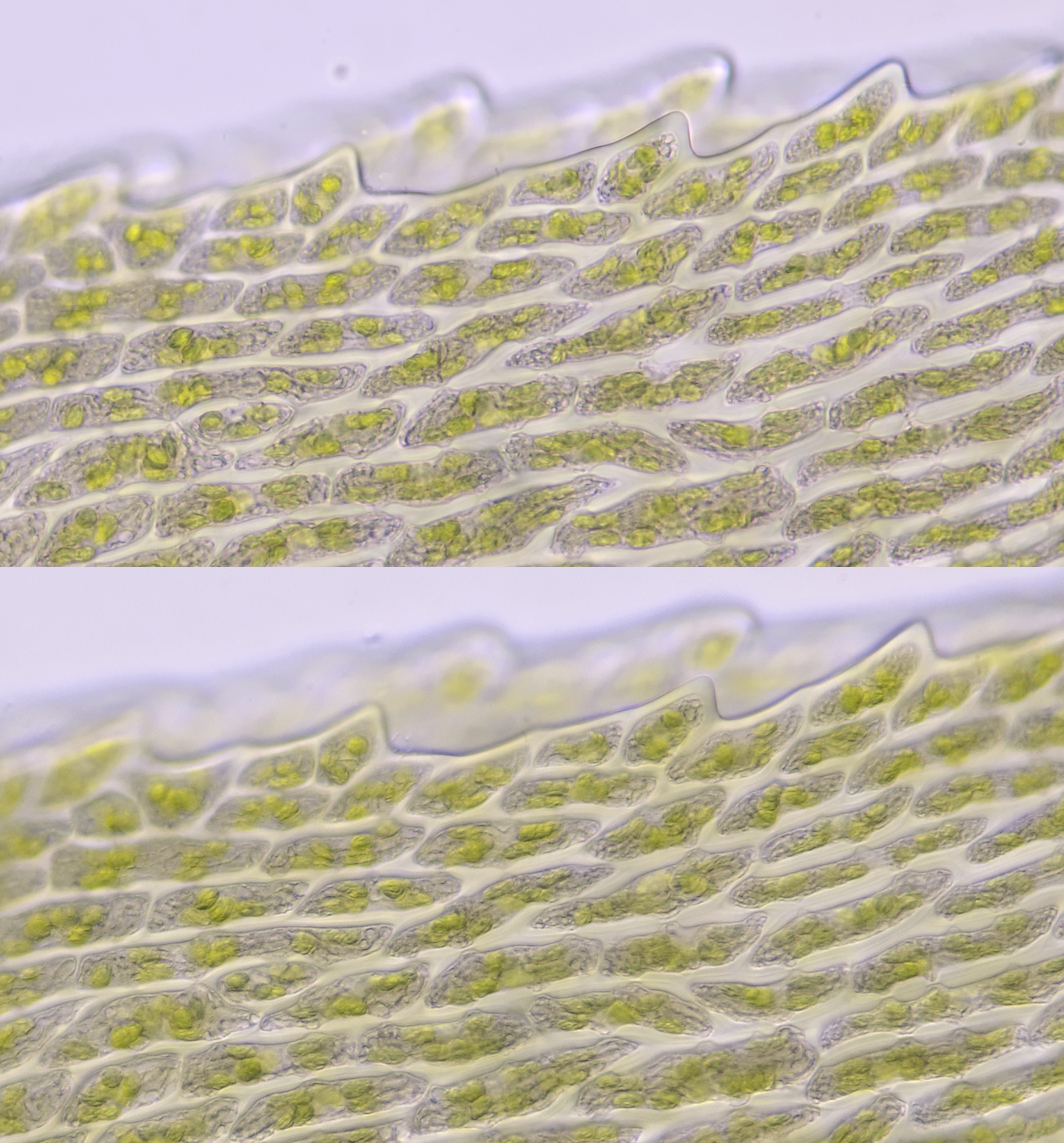
Detail van de bladrand van Dicranum uit Daun gefotografeerd met Olympus 40/0.65 in helderveld-belichting (boven) en schuine belichting (onder).
Dicranum, gevonden tijdens vakantie in Schotland. Macro-opname (links), micro-opnames met Olympus Plan 20/0.40 (midden) en met Olympus 40/0.65 (rechts). De foto's werden gemaakt met de Olympus HSA microscoop die ik tijdens vakantie had meegenomen.
Sphagnidae (veenmossen)
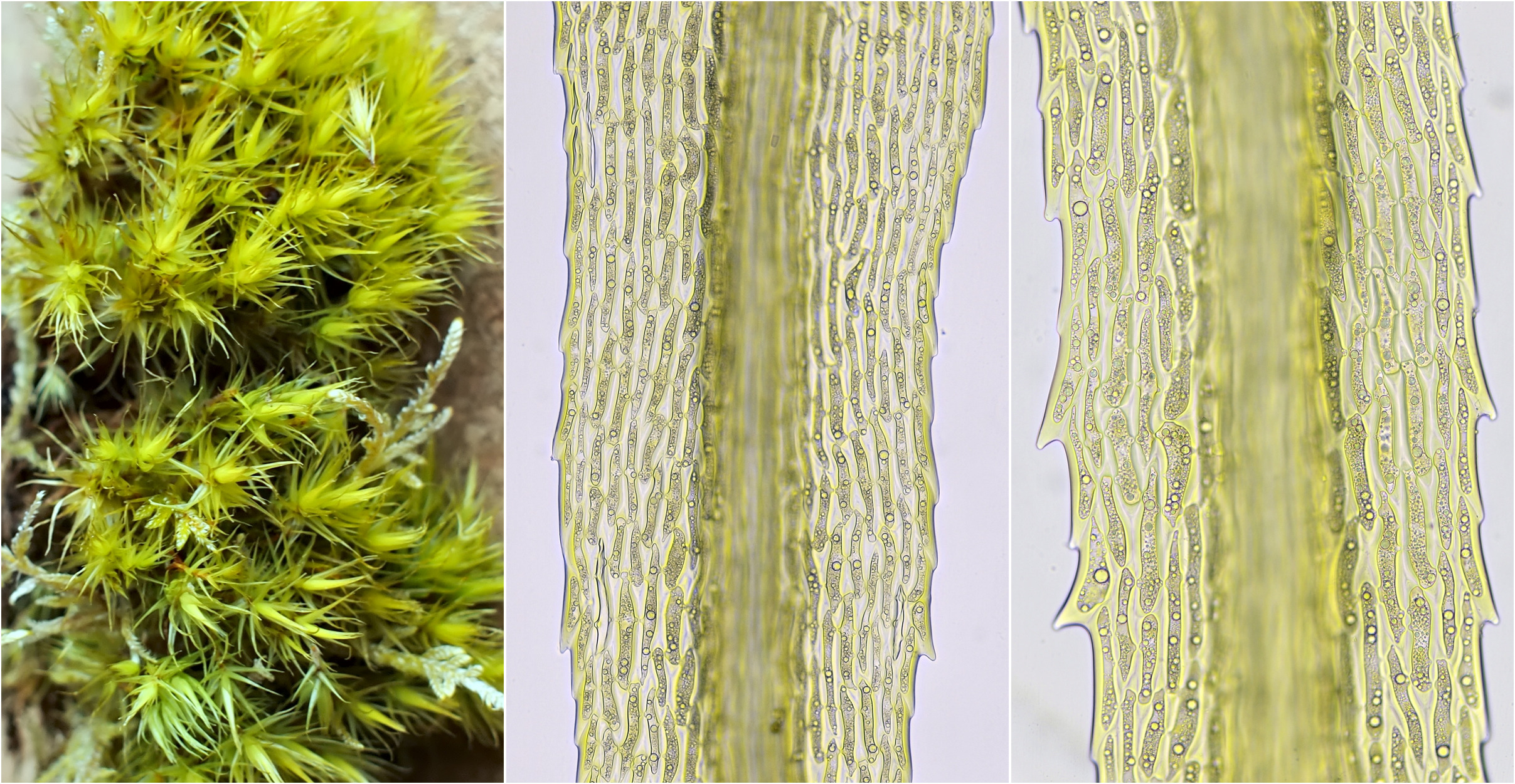
Tot de Sphagnidae of veenmossen behoort o.a. het geslacht Sphagnum. Dit mos komt in natte gebieden voor en de plant is met behulp van speciale cellen in staat om veel water vast te houden. Deze cellen, de zogenaamde hyaline cellen, functioneren als water-reservoirs. Het zijn dode cellen die door capillaire werking middels poriën water opnemen en daardoor als een soort spons werken. In het weefsel van de plant bevinden zich naast de hyaline cellen ook levende fotosynthetiserende cellen die een netwerk vormen.

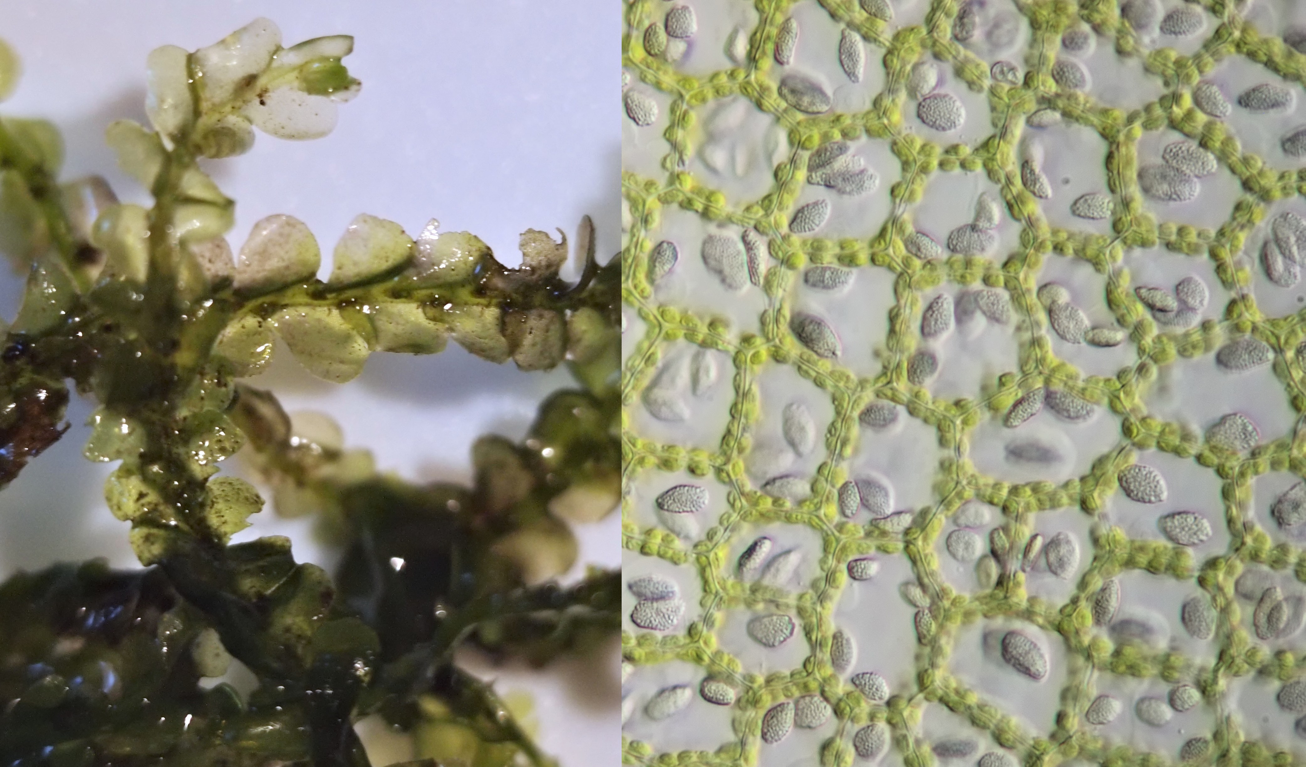
Een veenmos dat ik tijdens een wandelvakantie in Schotland tegenkwam (links). Rechts: opname gemaakt met de Olympus HSA microscoop die ik tijdens de vakantie bij me had. Objectief: Olympus Plan 20/0.40.
Detail-opname van het mos uit Schotland gemaakt met Olympus objectief 40/0.65. Hier zijn de hyaline cellen (transparant) goed te zien en ook de smalle levende cellen die een netwerk vormen.
Atrichum
Het geslacht Atrichum omvat de rimpelmossen en de soort Atrichum undulatum heeft duidelijk gerimpelde blaadjes met doorntjes aan de bladrand. De onderstaande foto's zijn van een mos dat ik aantrof tijdens een vakantie in Hornberg (Duitsland).
Blaadje van Atrichum gefotografeerd in schuine belichting met Carl Zeiss Objectief 10/0.22. Met een beetje fantasie doet dit denken aan een doolhof met boompjes waarin je gemakkelijk zou kunnen verdwalen..........
Detail-opnames van Atrichum gefotografeerd met Carl Zeiss Neofluar 40/0.75.
Thuidium
Thuidium of thuamos wordt o.a. gekenmerkt doordat het er als een mini-varen eruit ziet. Het wordt daarom ook wel varenmos genoemd.
Thuidium, gefotografeerd met Carl Zeiss objectief 10/0.22. Dit mos kwam ik tegen tijdens een vakantie in Hornberg (Duitsland).
Thuidium uit Hornberg (Duitsland) gefotografeerd met Carl Zeiss Neofluar 25/0.60.
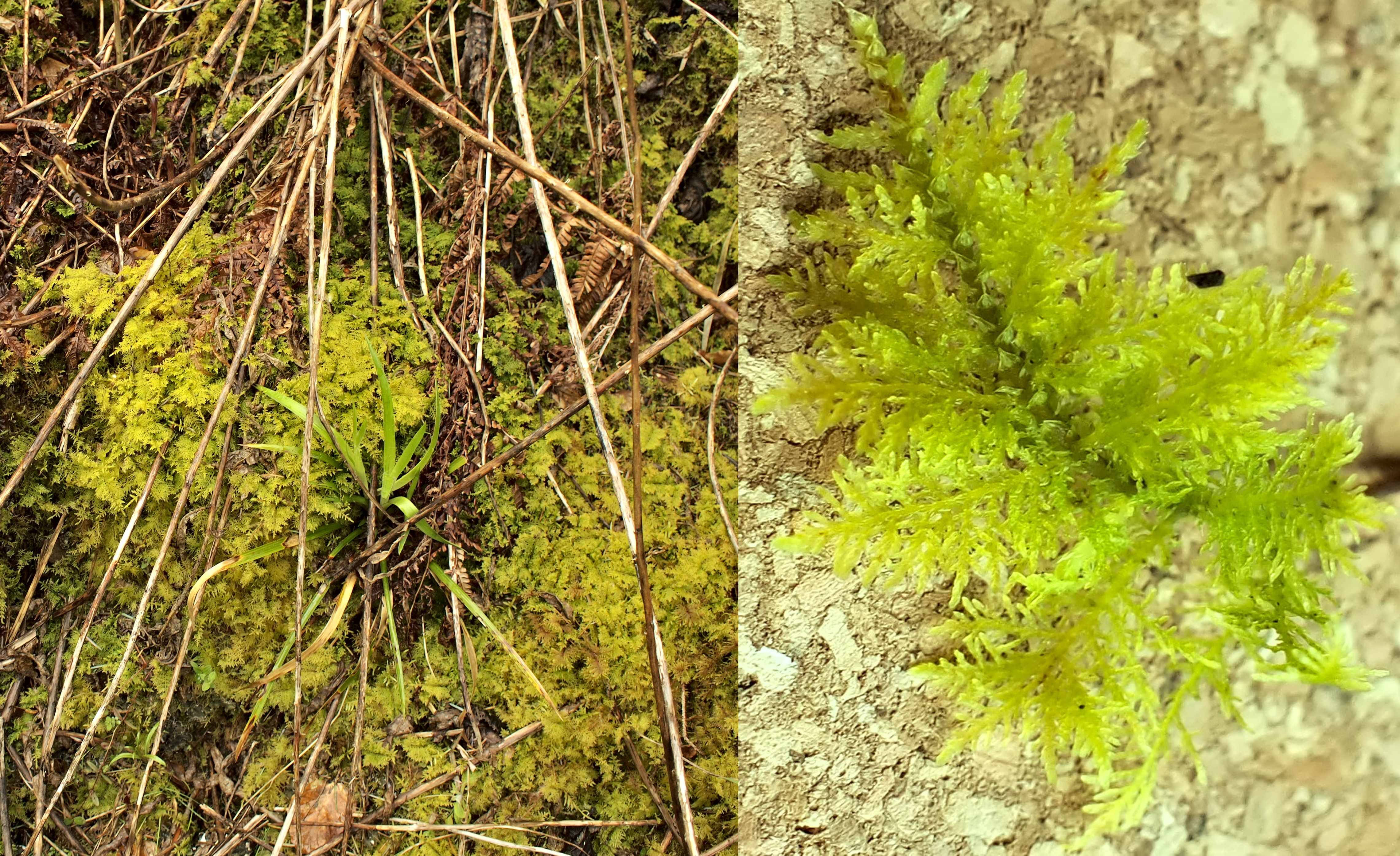
Habitatfoto en macro van Thuidium opgenomen tijdens een wandelvakantie in Schotland.
Thuidium uit Schotland gefotografeerd met Olympus 10/0.25 (links) en Olympus Plan 20/0.40 met de Olympus HSA microscoop die ik had meegenomen.
Thuidium, gevonden op de Utrechtse Heuvelrug. Gefotografeerd in helderveld (links) en donkerveld (rechts) met Carl Zeiss objectief 10/0.22.
Literatuur
Landwehr, J. (1966). Atlas van de Nederlandse bladmossen. Amsterdam: uitgave Koninklijke Nederlandse Natuurhistorische Vereniging.
Bold, H. C. (1973). Morphology of Plants. New York: Harper & Row, Publishers, Inc.