Introduction
When the water in ditches, ponds and puddles shows a green color, everyone knows that it is caused by algae. When green floating algae masses are present, they are usually filamentous green algae. Their characteristic feature is that they consist of long chains (filaments) of connected cells that at some point become intertwined and form a network. The cells have chloroplasts with shapes characteristic of the species. The group belongs to the Chlorophyta (green algae). During hot and sunny periods in the summer, large masses of algae can form on ditches and ponds. The ditch becomes suffocated and when the algae die, an anoxic environment arises. An alga called Spirogyra is usually the cause of this. This alga feels slimy and can be recognized by that alone. Other common filamentous algae include Cladophora and Oedogonium. It is interesting to study filamentous algae under the microscope, even though there can be occasional disappointment when it turns out that a sample taken contains Spirogyra for the umpteenth time…………
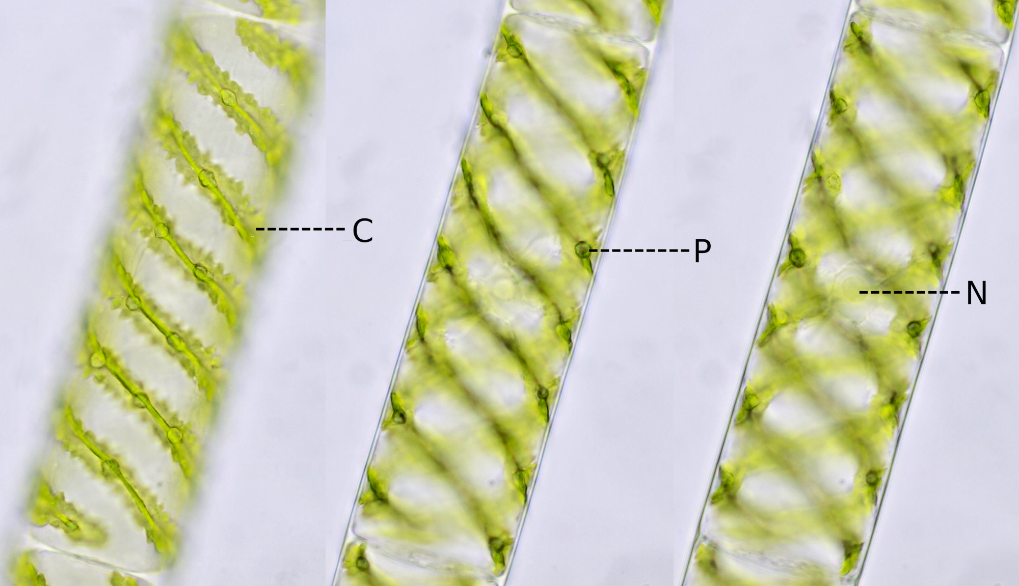
The chloroplasts of Chlorophyta are characterized by the presence of pyrenoids. These are round, thickened structures in which the enzyme RuBisCO is concentrated and where CO2 fixation takes place.
Spirogyra
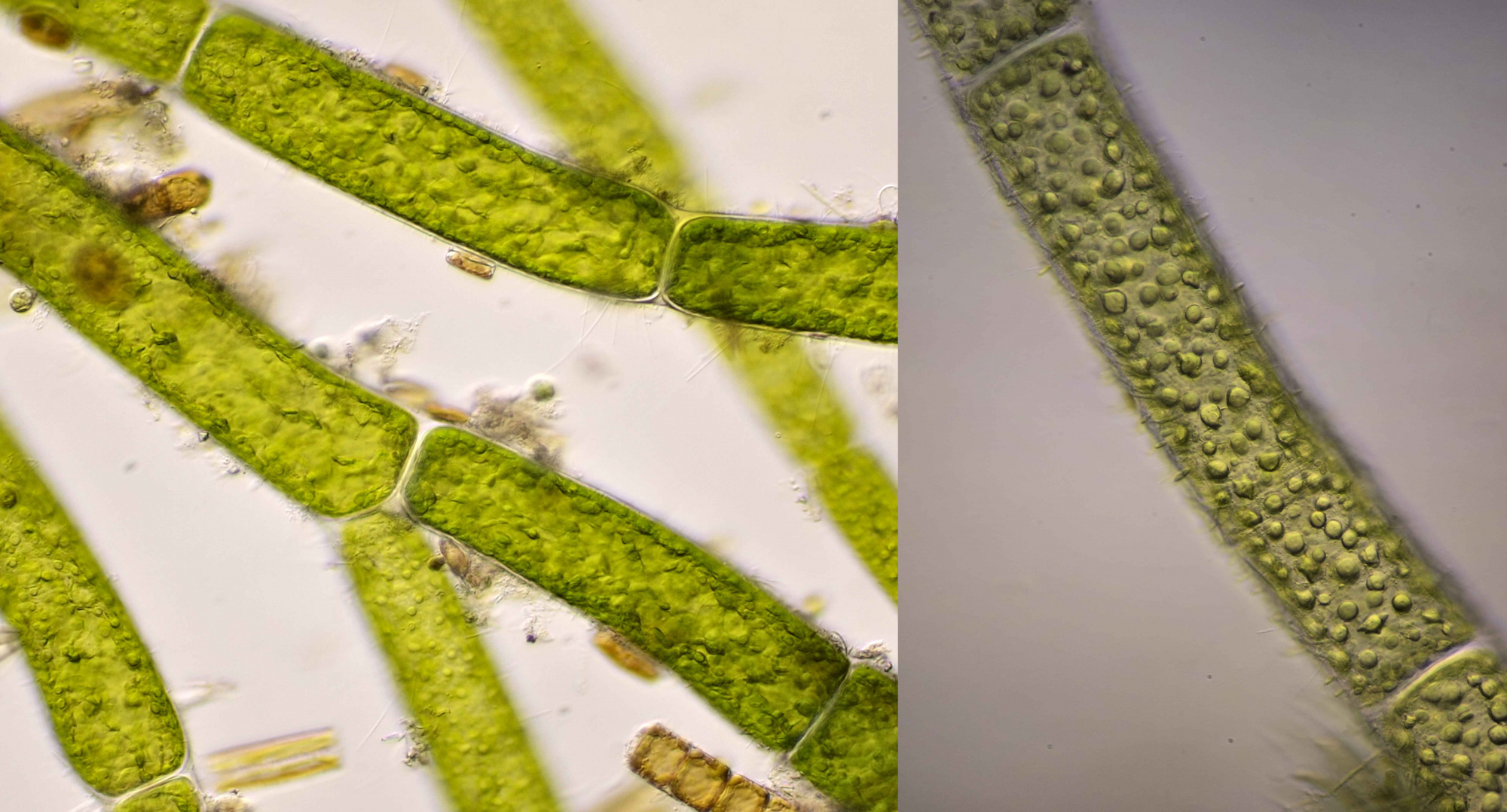
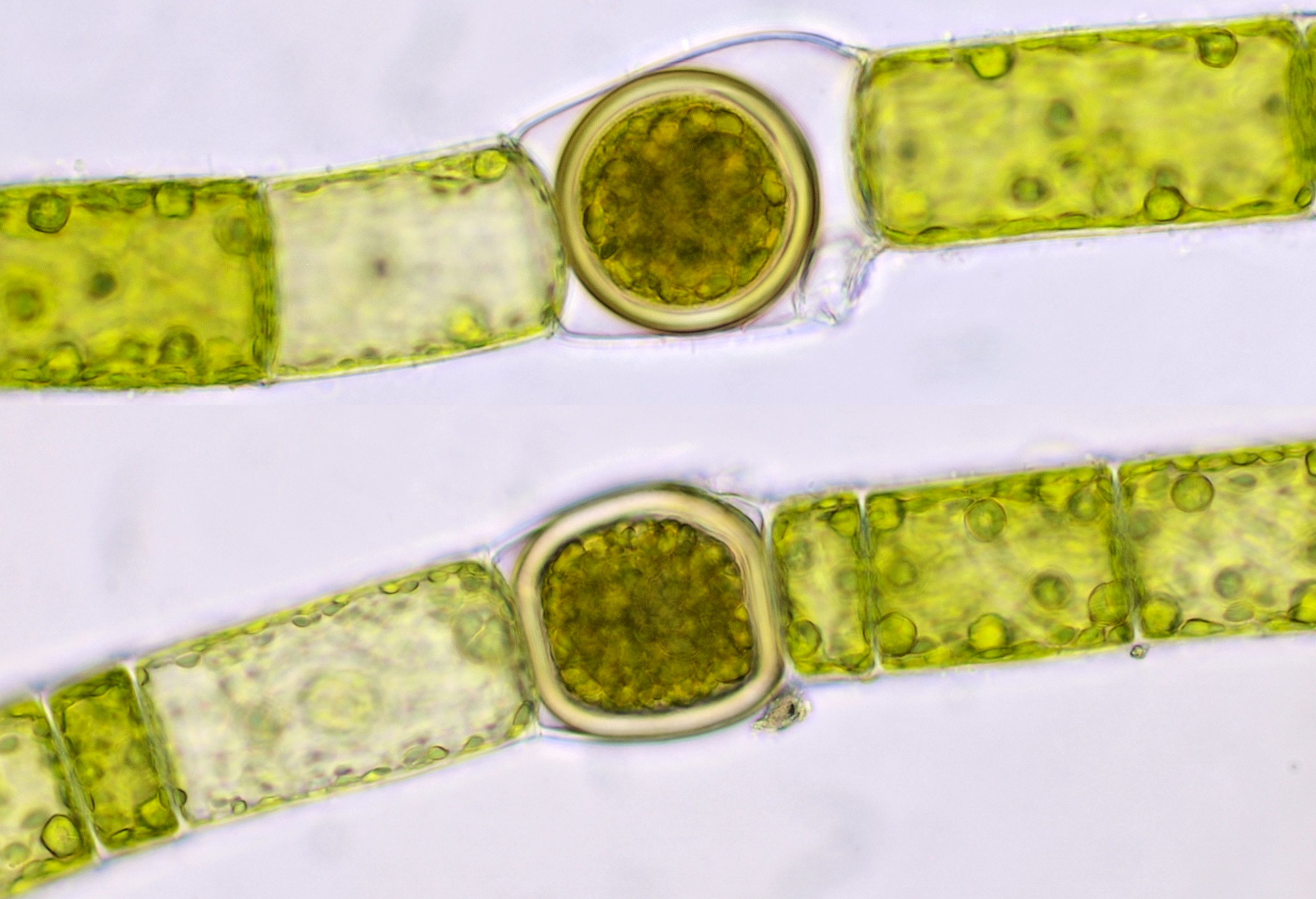
Spirogyra gets its name from the spiral-shaped chloroplasts in the cells. About 100 species of this algae occur in central Europe and it is sometimes called 'pond scum'. Sexual reproduction occurs by a process called conjugation in which two filaments become adjacent to each other and adjacent cells from the two strands fuse their cytoplasm (scalariform conjugation). The two cells form protrusions (papillae) that touch each other, after which the contents of one of the cells transfer to the other cell. This process can also take place between adjacent cells in a single filament (lateral conjugation). After the contents of both cells have fused, a zygote is created with a thick cell wall and a new filament is later formed from this zygote. Spirogyra forms long unbranched filaments that are surrounded by a gel-like sheat that makes the algae slimy to the touch. The diameter of the filaments varies from 8-170 μm, a considerable range from very thin to very thick filaments. Sometimes a cell nucleus can be seen and the pyrenoids are usually clearly visible in the spiral chloroplasts. The cells may contain a single or multiple chloroplasts. Spirogyra is a nice study object to observe cytoplasmic streaming (cyclosis).
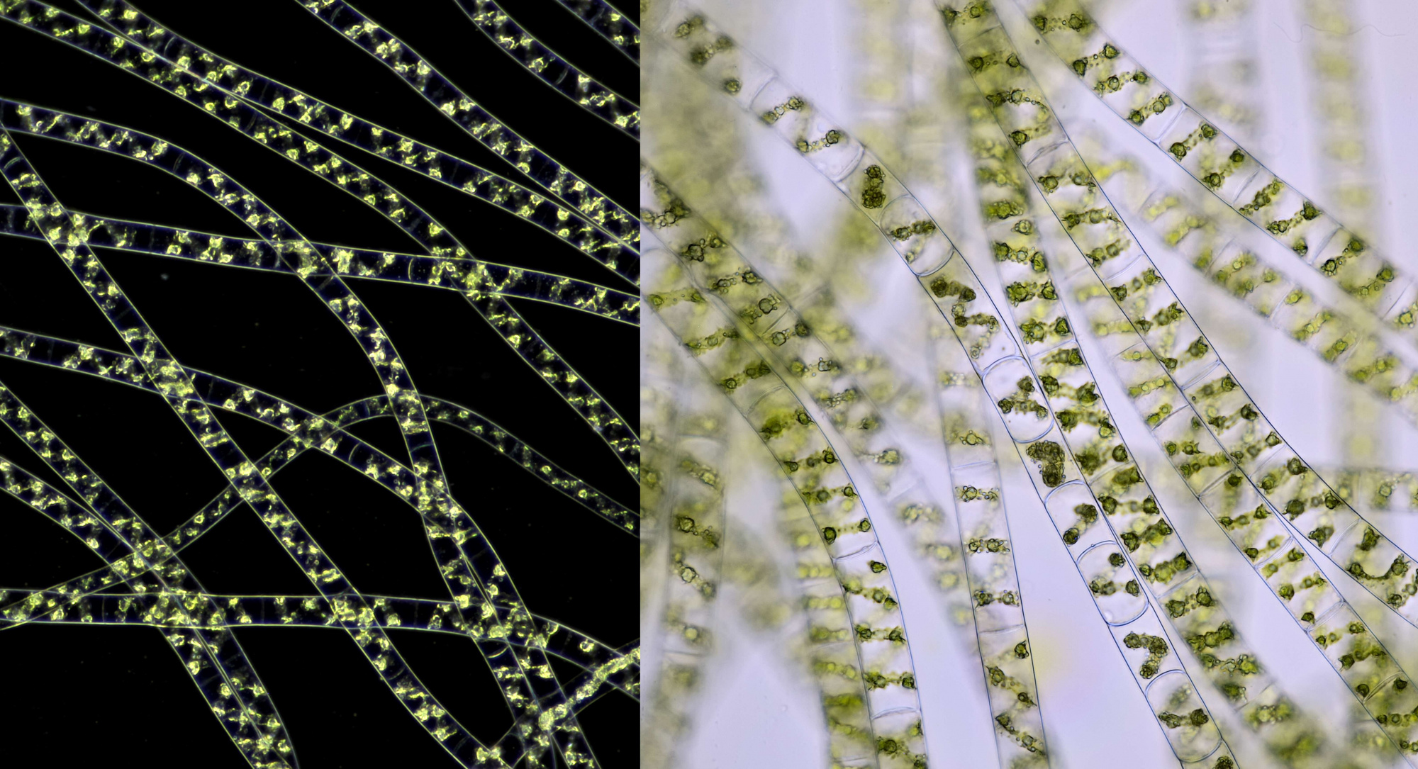
Spirogyra photographed with Carl Zeiss 10/0.22 in darkfield illumination (left) and normal brightfield with Carl Zeiss Jena Apo 16/0.40 (right).
A single cell of Spirogyra photographed in different focal planes to see the details more clearly. C: chloroplast. P: pyrenoid. N: nucleus. Objective: Zeiss-Winkel 40/0.65.
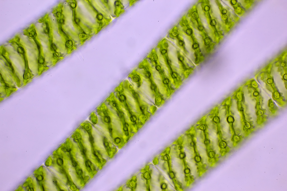
Spirogyra photographed in brightfield with Carl Zeiss Neofluar 40/0.75.
Spirogyra photographed with circular oblique illumination which gives a more spatial impression. Objective: with Leitz Pl Apo 25/0.65.
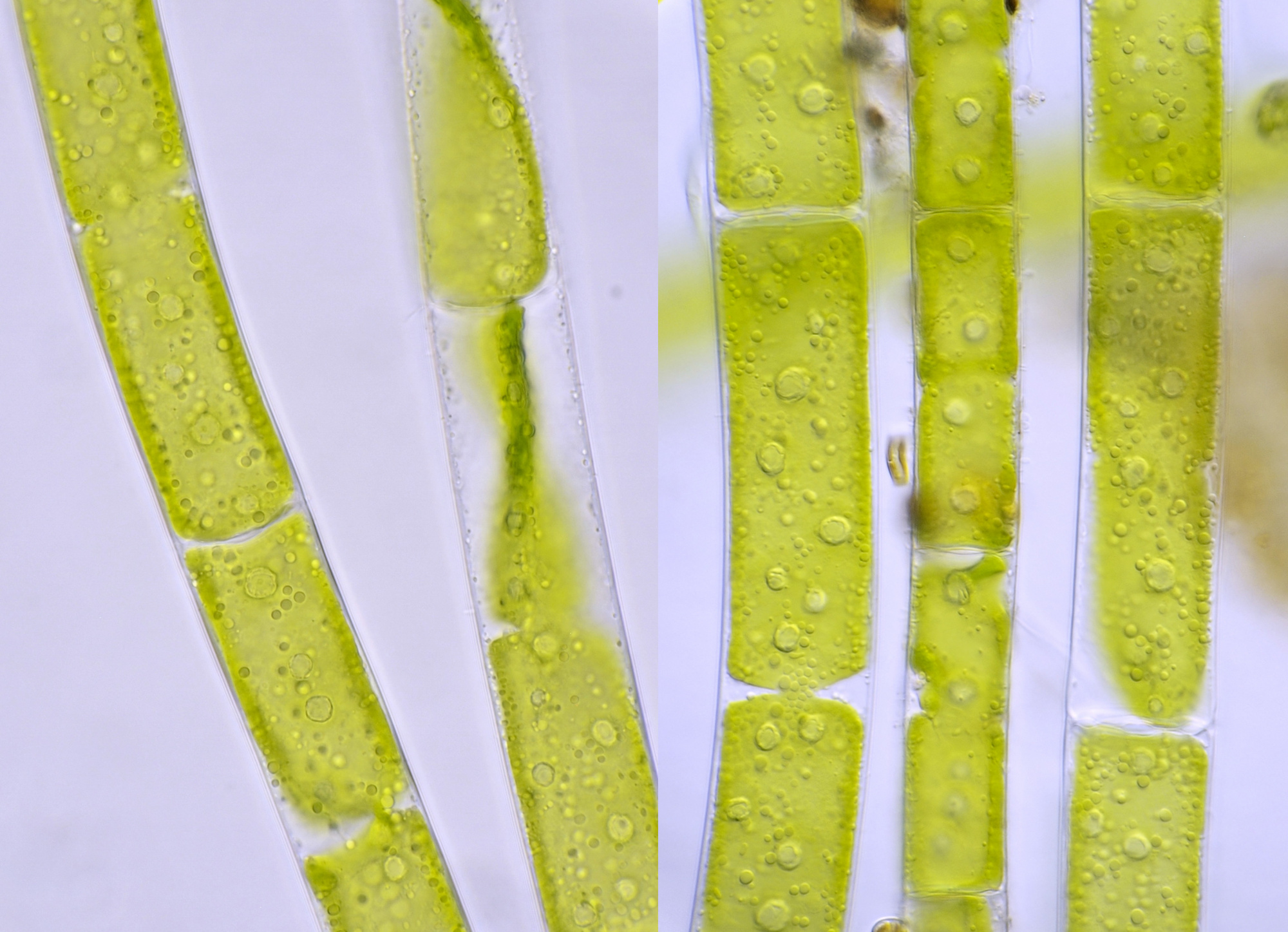
Spirogyra photographed with Carl Zeiss 63/0.80. Diameter of the filament is approx. 48 μm.
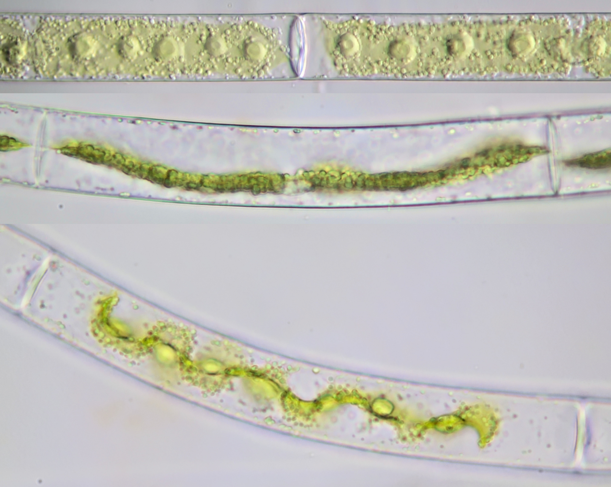
Very large Spirogyra specimen photographed with Olympus 10/0.25. The image shows atypical chloroplasts; this alga is dying and the chloroplasts are fragmenting.
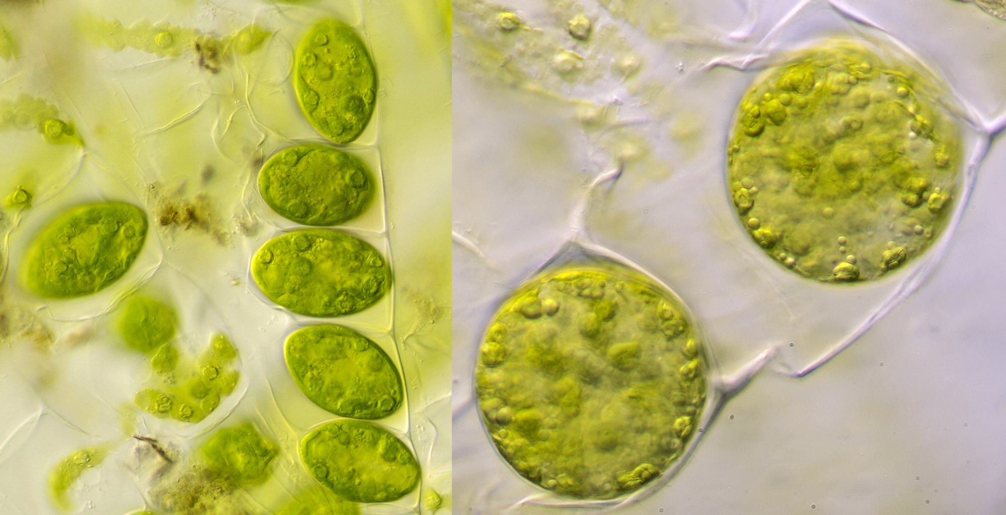
Zygotes of Spirogyra. Objective: Zeiss-Winkel 40/0.65.
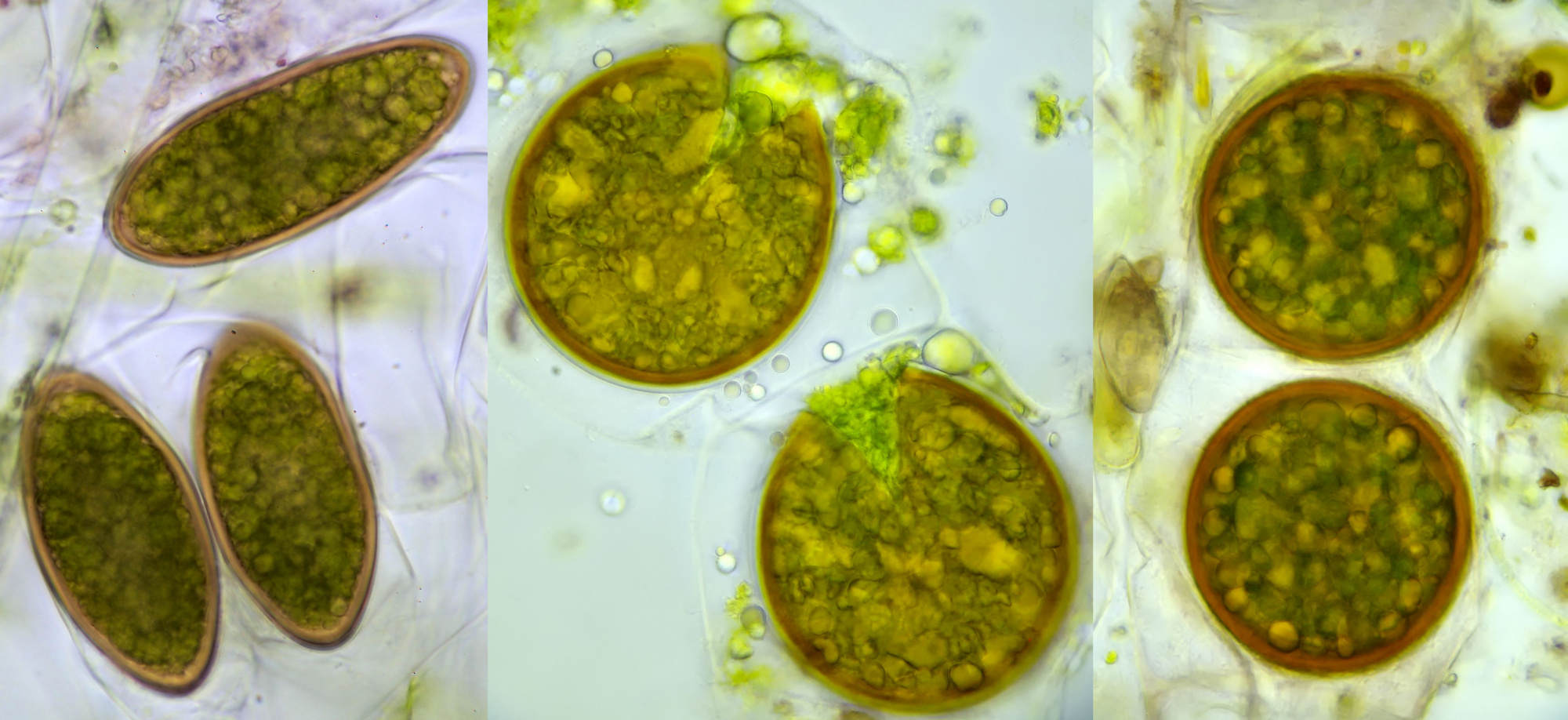
Zygotes of Spirogyra. Left with Carl Zeiss Neofluar 40/0.75, middle and right with Fl Oel 54/0.95.
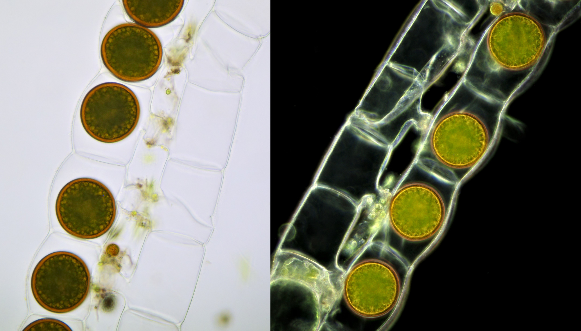
Zygotes of Spirogyra after conjugation has completed. The protrusions between the two filaments are still visible. Photographed with Leitz Pl Apo 25/0.65 in normal brightfield (left) and darkfield illumination (right). Camera: Olympus Stylus 725 SW.
Cytoplasmic streaming (cyclosis) in Spirogyra. Objective: Leitz Pl Apo 40/0.75.
Zygnema
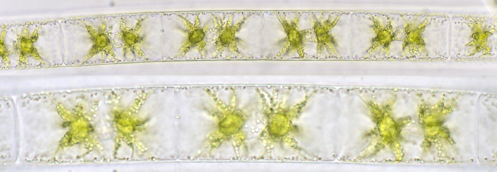
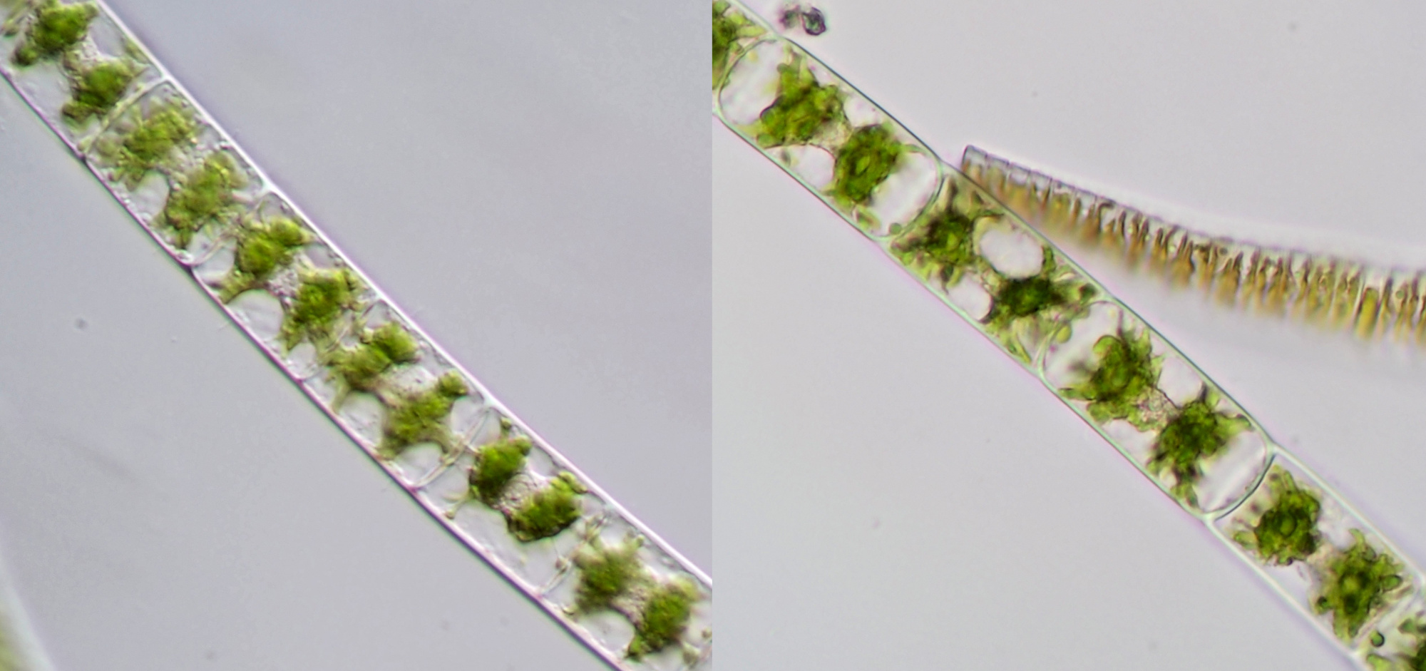
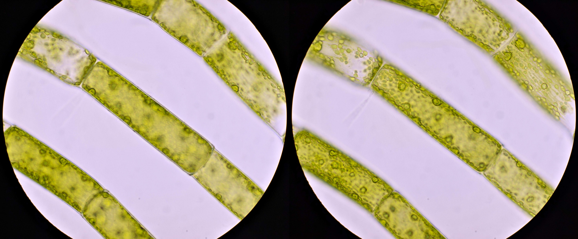
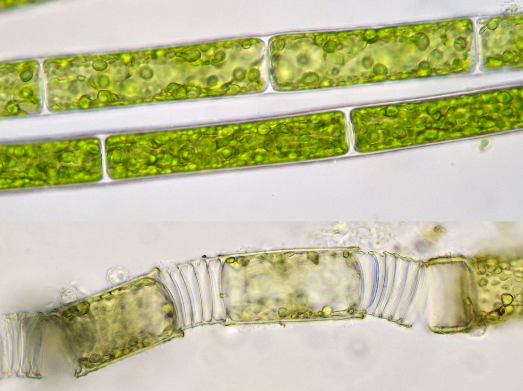
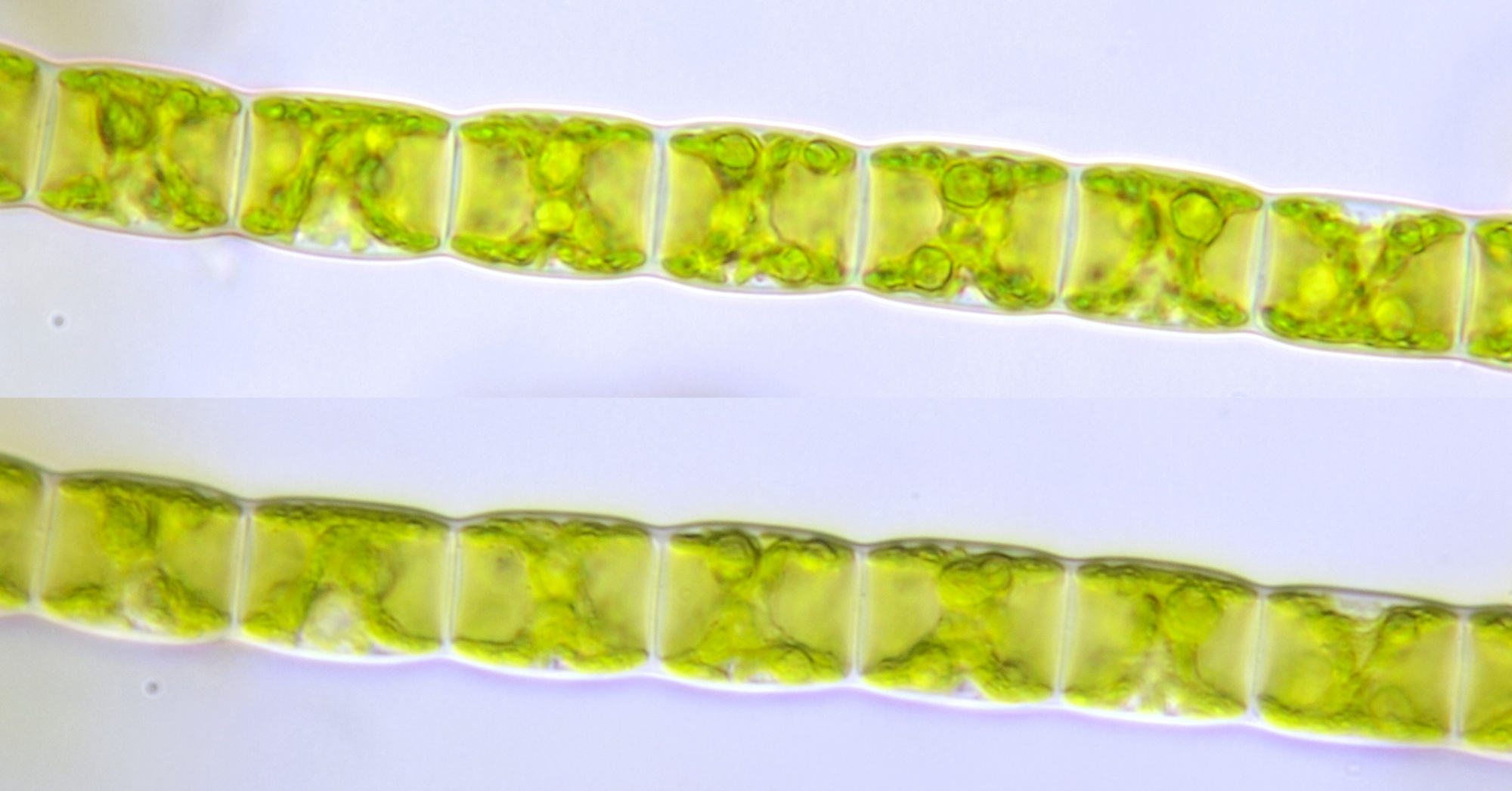
An alga that strongly resembles Spirogyra is Zygnema. Like Spirogyra, this alga belongs to the conjugating algae (Conjugatophyceae) and only occurs in fresh water. Each cell contains two stellate chloroplasts, which makes it easy to distinguish the Zygnema from Spirogyra. The diameter of the cells varies from 20-40 μm.
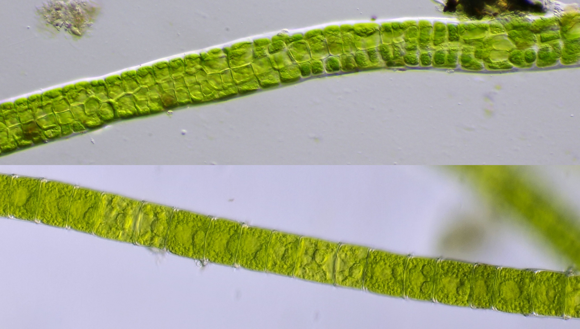
Zygnema, photographed with Olympus 40/0.65 (upper image) and WI 63/0.85 (lower image).
Zygnema, photographed with oblique illumination (left) and normal brightfield (right) during holiday in Hornberg, Schwarzwald. Objective: Carl Zeiss Neofluar 25/0.60.
Cladophora
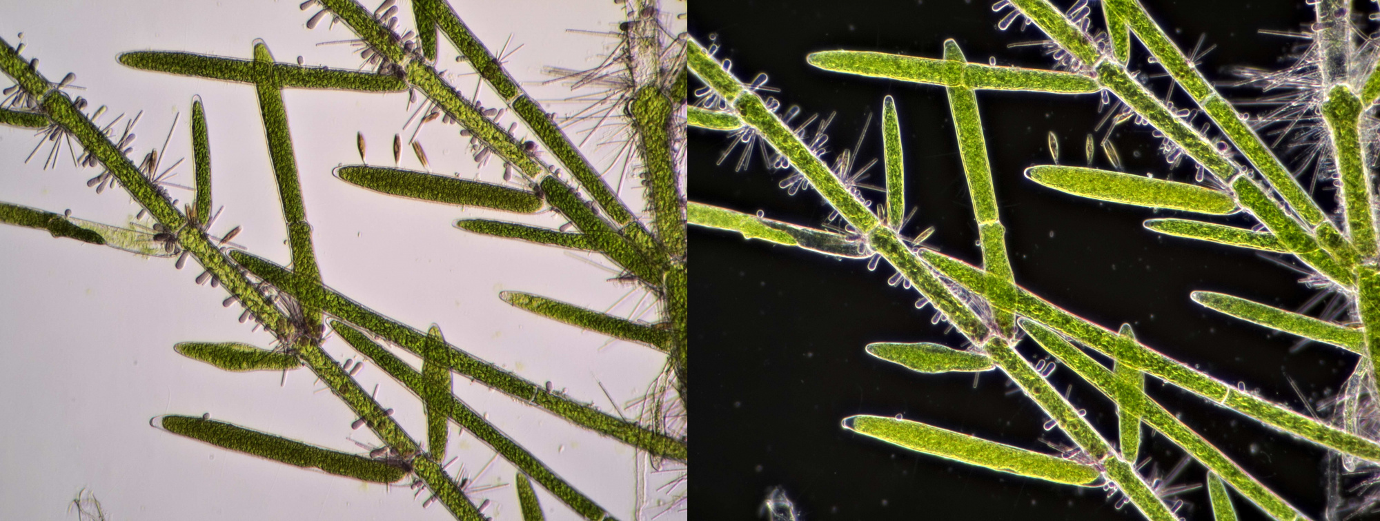
Cladophora is widely distributed in both fresh and salt water. This filamentous algae, 10 – 200 μm in diameter, forms branches and feels rough to the touch when a sample is removed from the water. The cells have multiple nuclei and the chloroplast consists of irregular segments or a complete network, the latter especially in younger cells. Both sexual and asexual reproduction take place. Asexual reproduction takes place by zoospores with 4 flagella and these are formed in the cells at the ends of the chains. Cladophora is often overgrown with other organisms such as diatoms and cyanobacteria.
Cladophora, from a tropical aquarium, photographed with oblique lighting (left) and darkfield illumination (right). Objective: Zeiss F10/0.25.
Cladophora, from a tropical aquarium, photographed with Leitz NPL Fluotar 25/0.55.
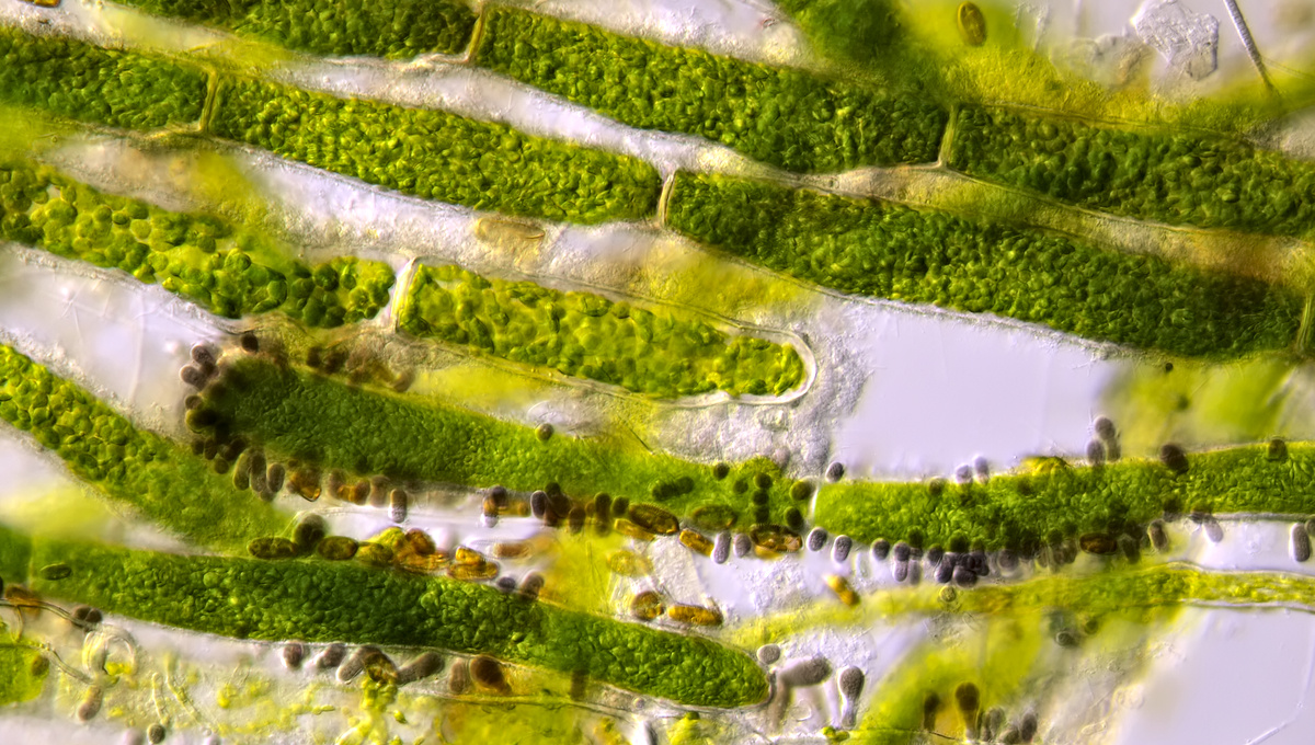
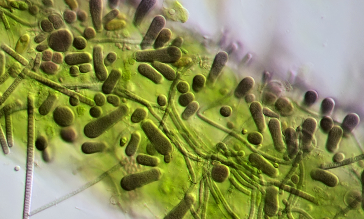
Cladophora, from a tropical aquarium, completely overgrown with different species of cyanobacteria. Objective: Carl Zeiss Neofluar 25/0.60.
Cladophora from the Maas near Kessel (left, Zeiss-Winkel 40/0.65) and from my pond (right, Zeiss-Winkel 25/0.45).
Oedogonium
Oedogonium is a common filamentous alga that does not form branches. The diameter of the filaments varies from 3-60 μm and the cells contain highly branched, reticulate chloroplasts. This alga often grows epiphytically on other algae or aquatic plants. Some other algae have a similar appearance, but there is still a characteristic specific to Oedogonium: the so-called cap cells. These are the remains of cell divisions.
Reproduction occurs both sexually and asexually. In the latter case, zoosporangia surrounded by a thick wall are formed from which motile zoospores develop. After a while, these will leave the cell, moving with the help of flagella. Eventually, a new filament will develop from a zoospore.
Filaments of Oedogonium photographed at different focal planes: Objective: Zeiss-Winkel 40/0.65.
Oedogonium photographed with Carl Zeiss Neofluar 40/0.75 (upper image). The lower photo shows the cap cells, photographed with Zeiss-Winkel 40/0.65.
Filaments containing a zoösporangium. Objective: Zeiss-Winkel 40/0.65.
Mougeotia
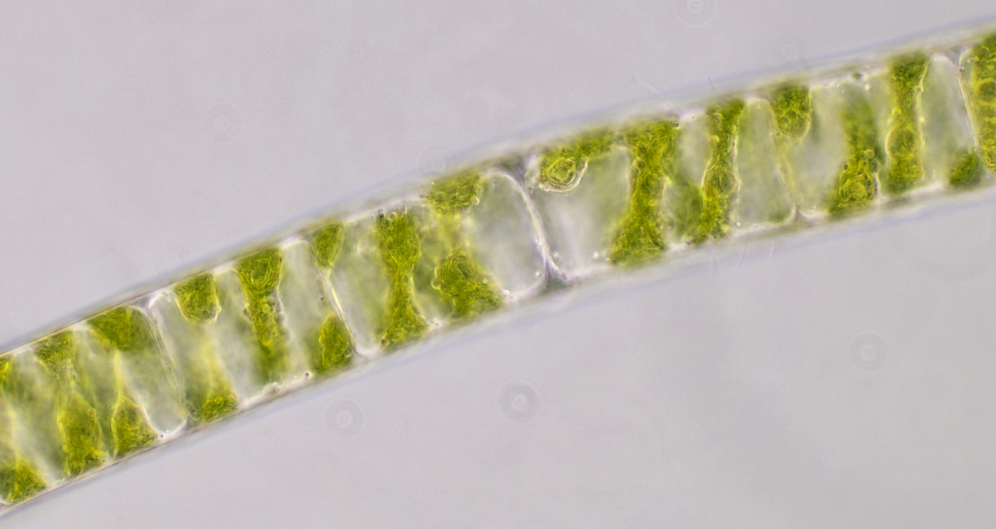
A filamentous alga that can have a diameter of 4-40 μm. This alga has the special feature that the chloroplast can adapt to the light intensity. In weak light, the flat side (largest surface) of the chloroplast is turned towards the light.
Different specimens of Mougeotia. Above: appearance in dim light. Middle and below: the narrow side of the chloroplast in high light intensity. Objective: Zeiss-Winkel 40/0.65.
Mougeotia photographed with Carl Zeiss Apo 40/1.0
Ulothrix
Ulothrix (Ulothrix Kützing 1833) is a common unbranched filamentous alga that can be found in both fresh and salt water. Most species occur in fresh water and the filaments have a diameter of 5 – 70 μm. The cells located between the two ends of the chain (the intercalary cells) are usually as wide or wider than they are long. The filaments are connected to a surface (holdfast) via a basal cell at one of the ends. The cell at the other end is called the apical cell and has a rounded shape. The cells have a single chloroplast that has the shape of a bracelet. There are one or more pyrenoids present in the chloroplast.
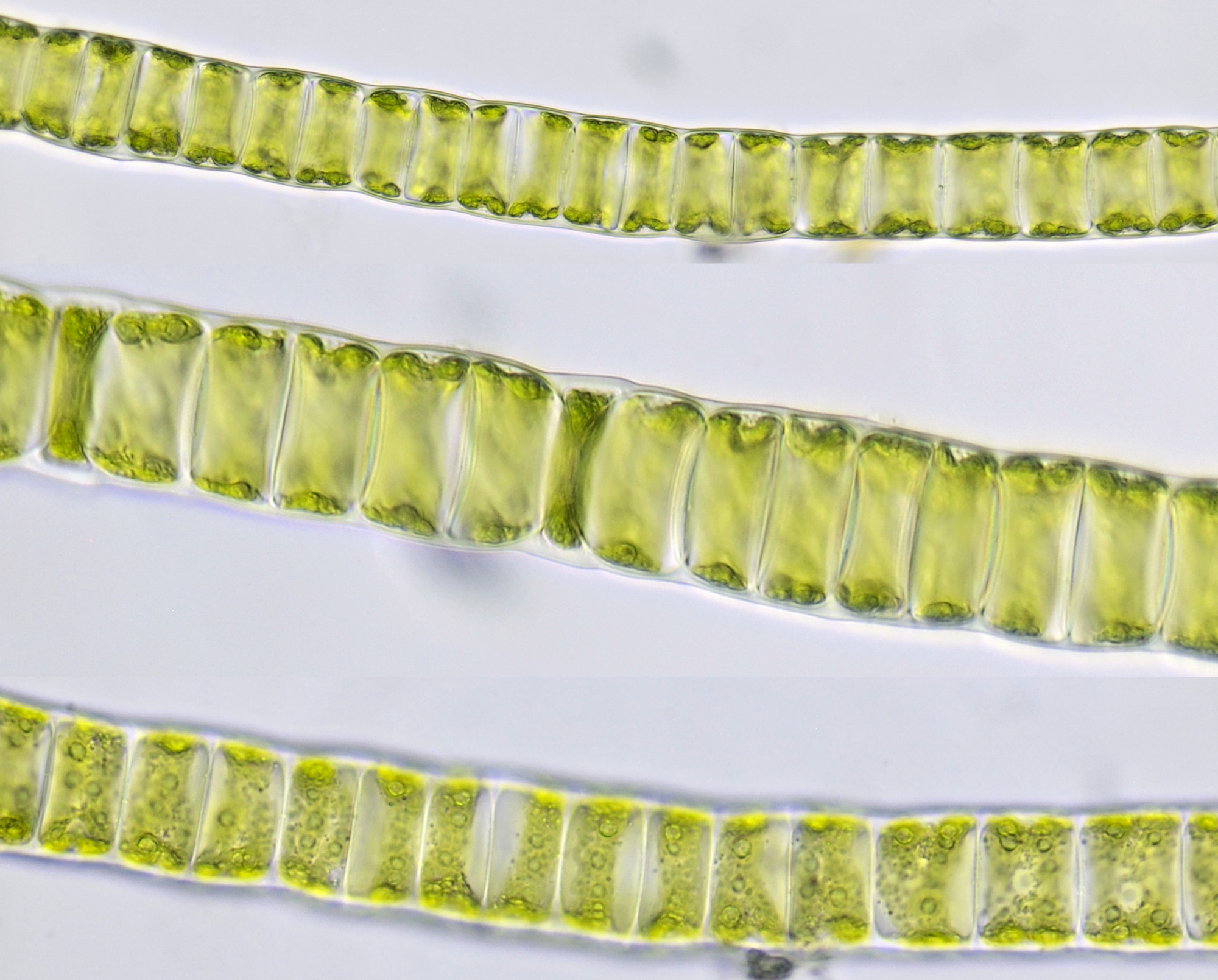
Ulothrix from the Mondsee (Austria), photographed with different exposures and focus. The pictures were taken with the Olympus HSA microscope that I took with me on holiday. Objective: Olympus 40/0.65.
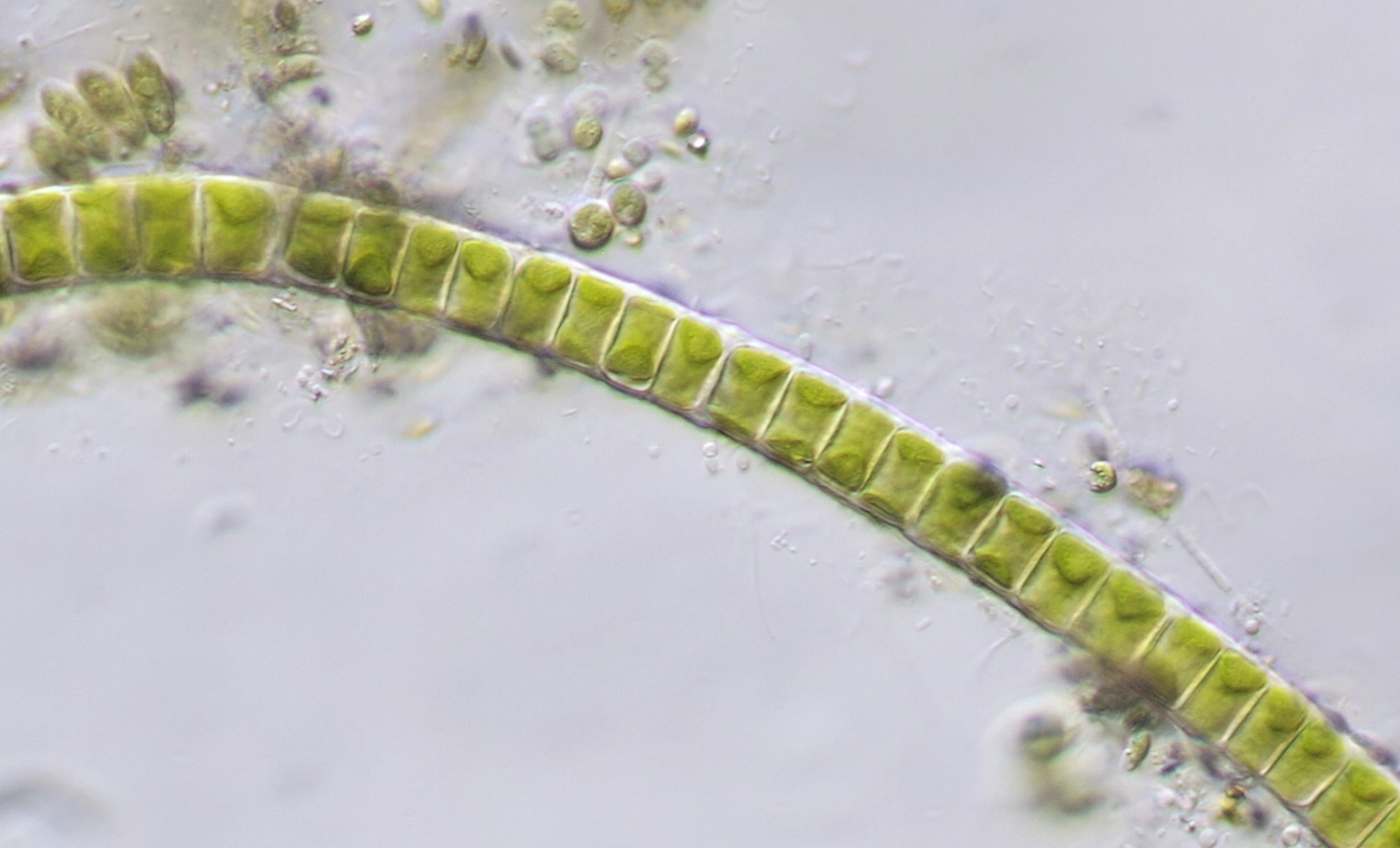
Ulothrix photographed with Zeiss-Winkel 40/0.65 in oblique illumination.
Chaetophora
Chaetophora is a branched filamentous alga that occurs attached to a substrate in flowing fresh waters. The diameter of the filaments varies from 4 - 10 μm and they are embedded in a gelatinous mass.
Chaetophora from the Tasbeek in Kessel (Limburg, Netherlands). Objective: Zeiss-Winkel 40/0.65.
Unknown algae
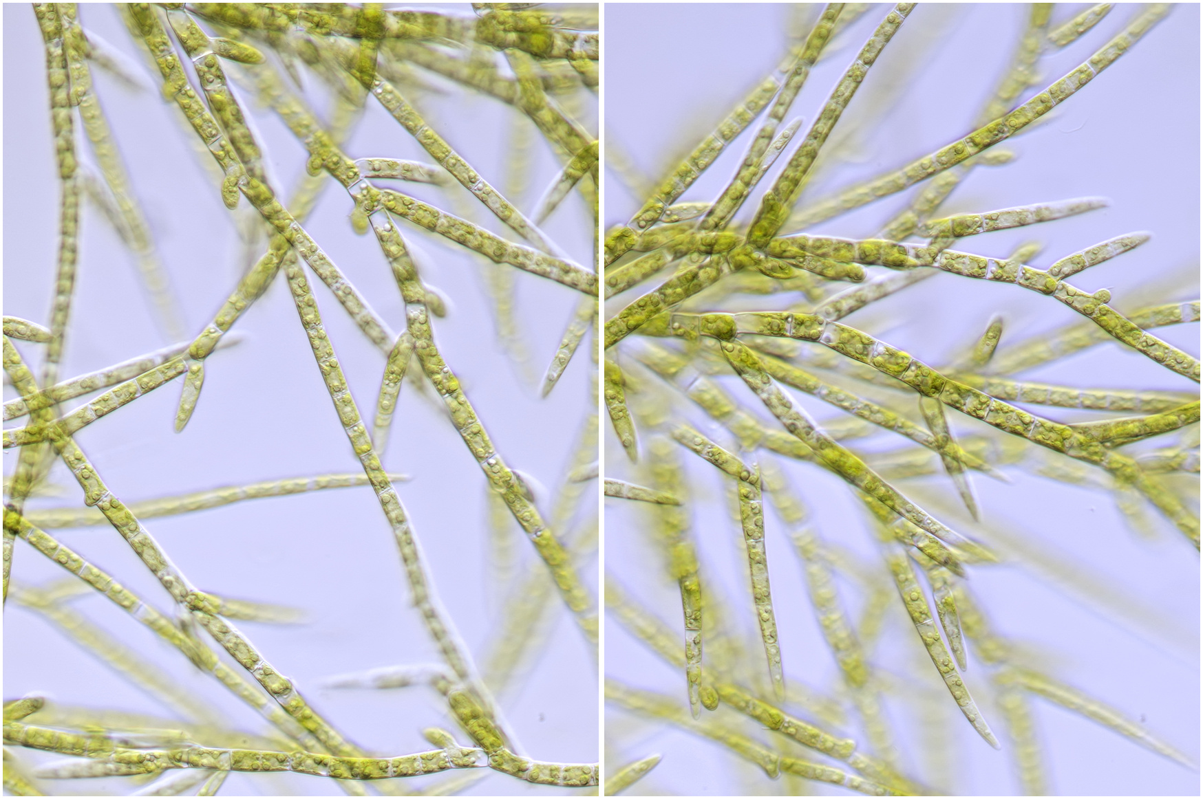
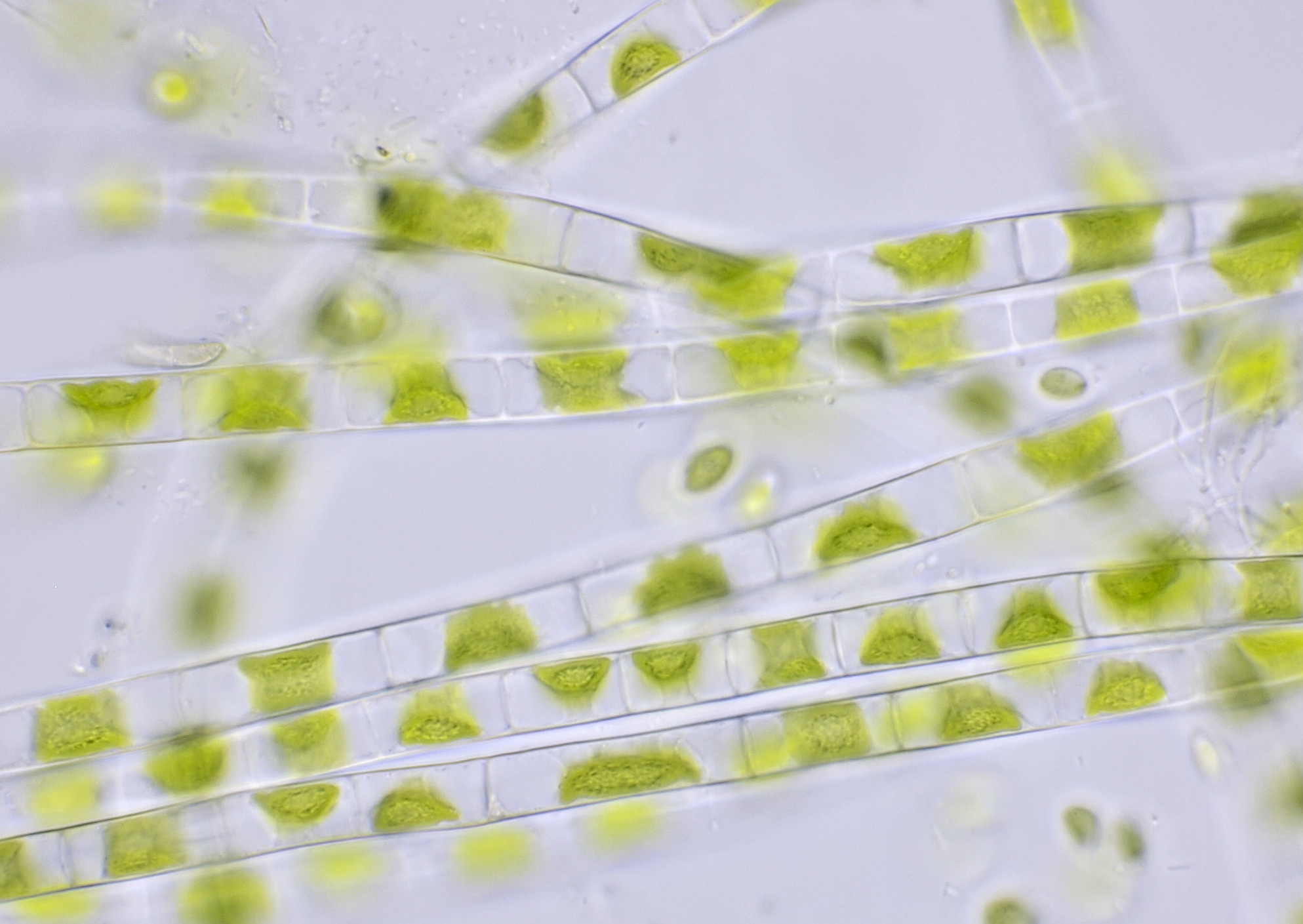
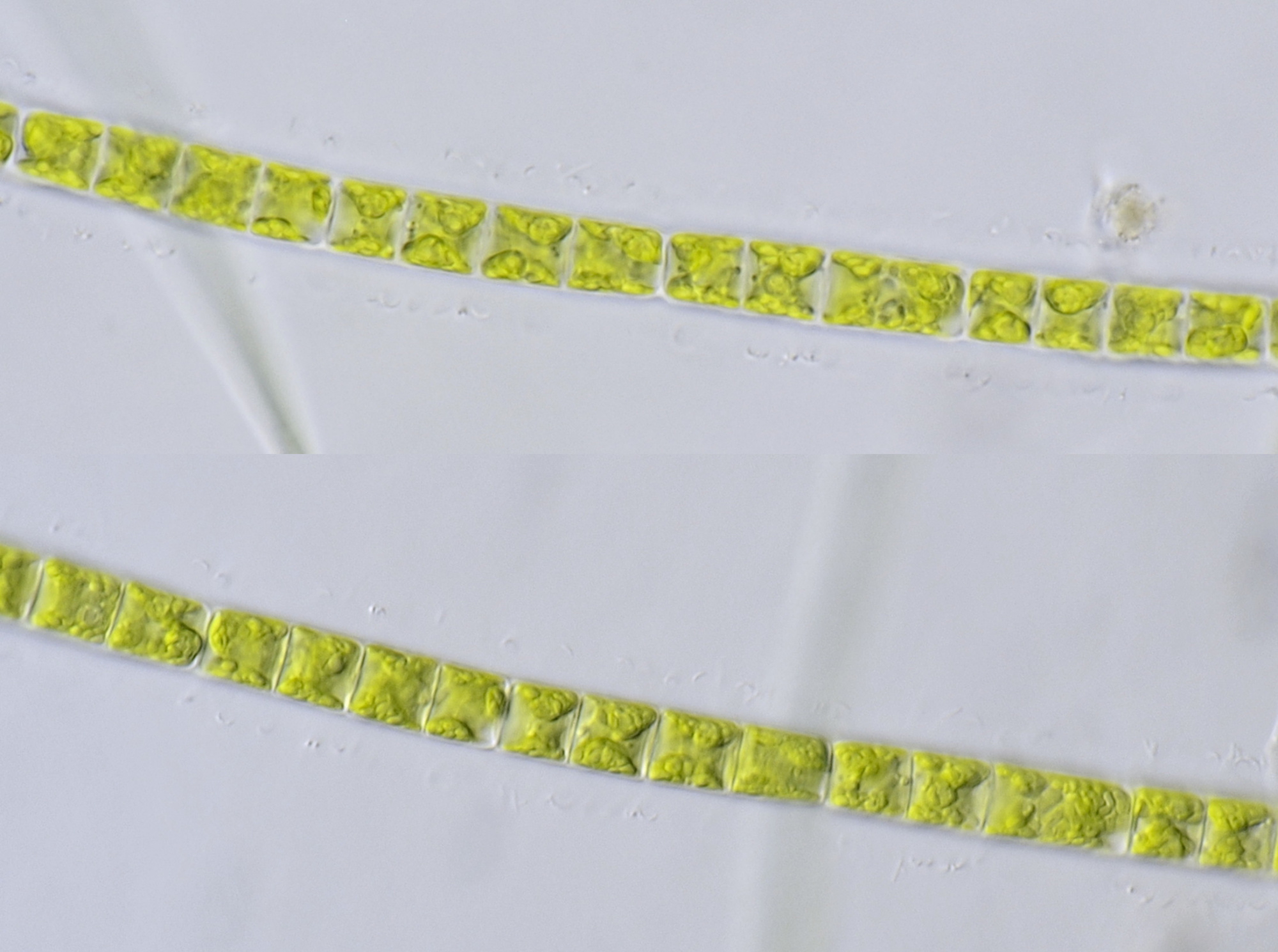
I have not yet been able to name certain filamentous algae that I encountered, hopefully that will come at a later stage. What follows is a selection of algae that I found at various locations in the Netherlands and abroad.
Algae from the green deposit on my balcony. Objective: Carl Zeiss Apo 40/1.0.
Alga from the river Maas in Kessel (Limburg, Netherlands) photographed in brightfield (above) and oblique illumination (below). Objective: Zeiss-Winkel 40/0.65.
A filamentous alga surrounded by a barely visible slime layer. Photographed in brightfield (top) and oblique illumination (bottom) with Carl Zeiss Apo 40/1.0.
Above: Filamentous alga from a ditch in Nieuw-Vredelust garden park, objective Carl Zeiss Plan 25/0.45. Below: Alga from the water in 't Loo park in Voorburg, objective Leitz Planapo 25/0.65. Both photos were taken with oblique lighting.
An algae I found in a bucket of water in the garden. Photographed with the Olympus HSA microscope and an F40/0.65 objective.
Literature
Linne von Berg, KH., Hoef-Emden, K., Marin, B., Melkonian, M. (2012). Der Kosmos Algenführer. Stuttgart: Franckh-Kosmos-Verlags-GmbH & Co.
Bold, H. C. (1973). Morphology of Plants. New York: Harper & Row, Publishers, Inc.
