Introduction
Diatoms are brown microalgae that can be found in all freshwater and marine environments. They are an important link in the ecological cycle. In streams and rivers you often see stones covered with a brown layer that is formed by diatoms. All algae have chloroplasts in which photosynthesis takes place. They perform the same function as the chloroplasts in the cells of terrestrial plants. The chloroplasts of diatoms have a brown color because, in addition to chlorophyll, they also contain the brown pigment fucoxanthin. Fucoxanthin is also found in brown seeweed that is common on beaches.
The cells of diatoms are surrounded by a two-part exoskeleton called a frustule that is made of silicon dioxide (the mineral in quartz). The frustule consists of two halves called valves that perfectly fit together. Depending on the symmetry of these valves, a distinction is made between centric (with radial symmetry) and pennate (with bilateral symmetry) diatoms. The frustules often have beautiful symmetrical shapes and refined structures. Diatoms can tell a lot about the water quality in lakes and rivers; they are good indicator organisms.
Diatoms are also used in human industry. An example of this is diatomaceous earth, a product that consists of the skeletons of diatoms and it is used in filter and purification systems.
Shown below is a small collection of pictures of diatoms that I found in water samples from various ditches and ponds.
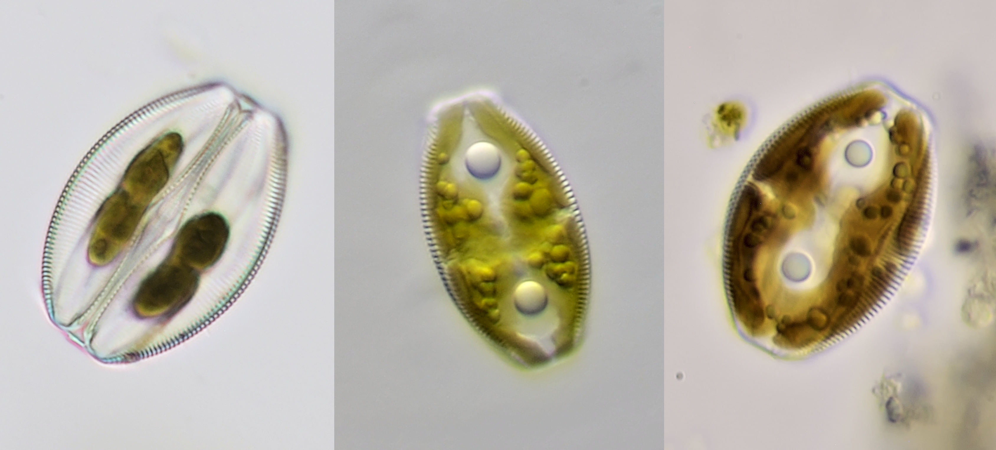
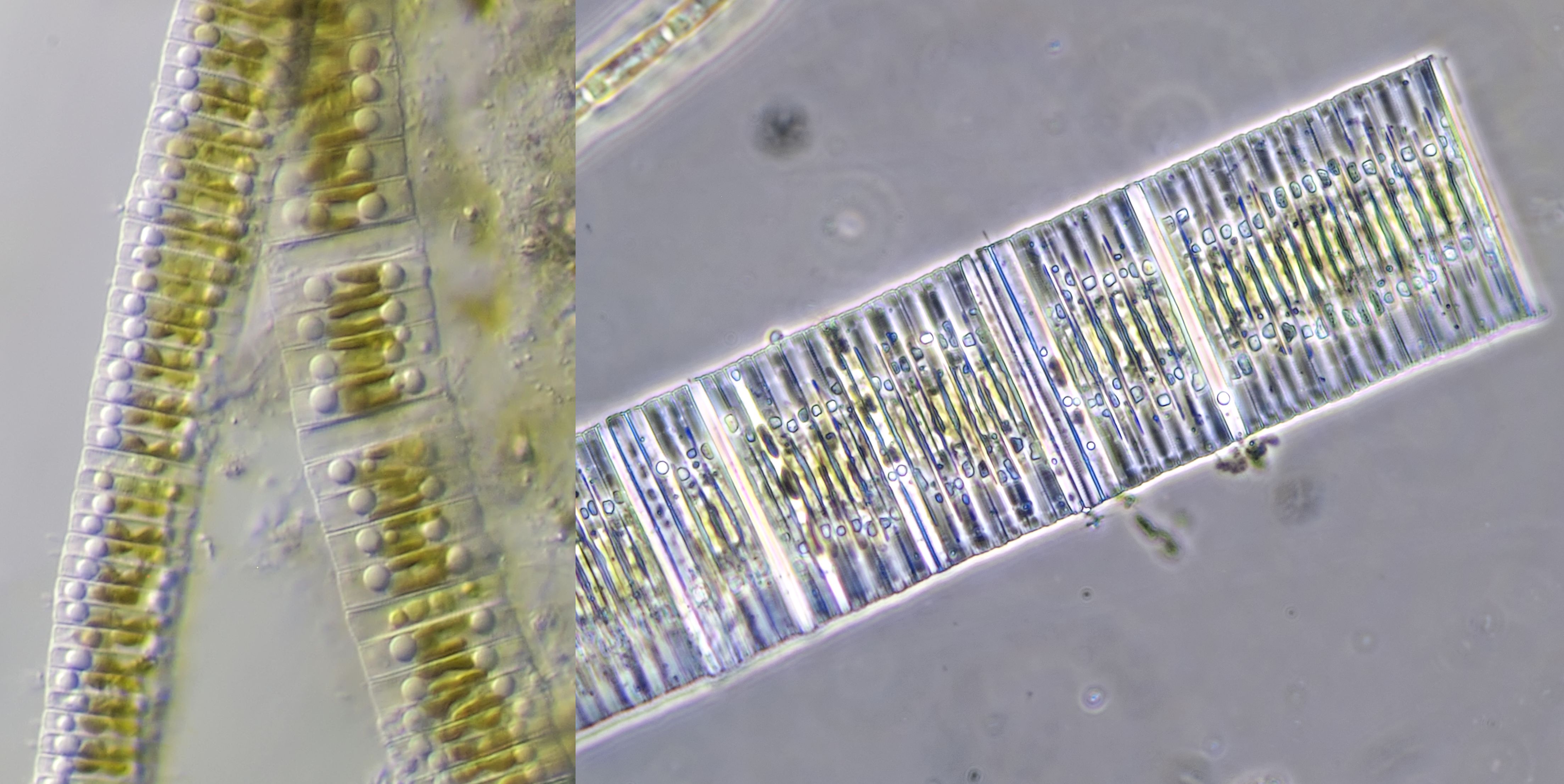
Diatoma
Diatoma is common in fresh and brackish water and is often notable for the characteristic zig-zag arrangement of the cells.
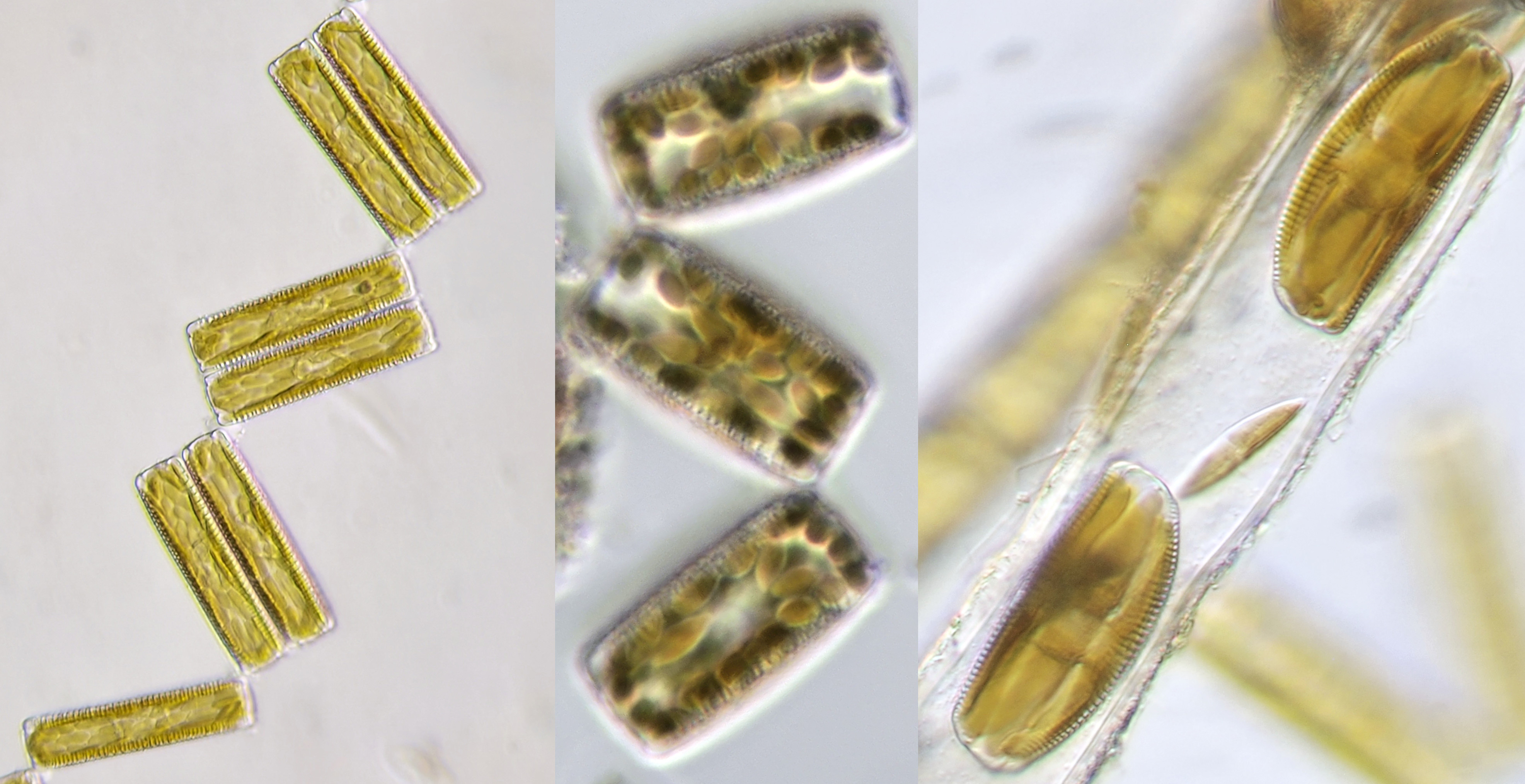
Diatoma, photographed with Carl Zeiss Neofluar 25/0.60 (left), Leitz Pl Apo 25/0.65 with circular oblique illumination (center) and Zeiss-Winkel 40/0.65 (right).
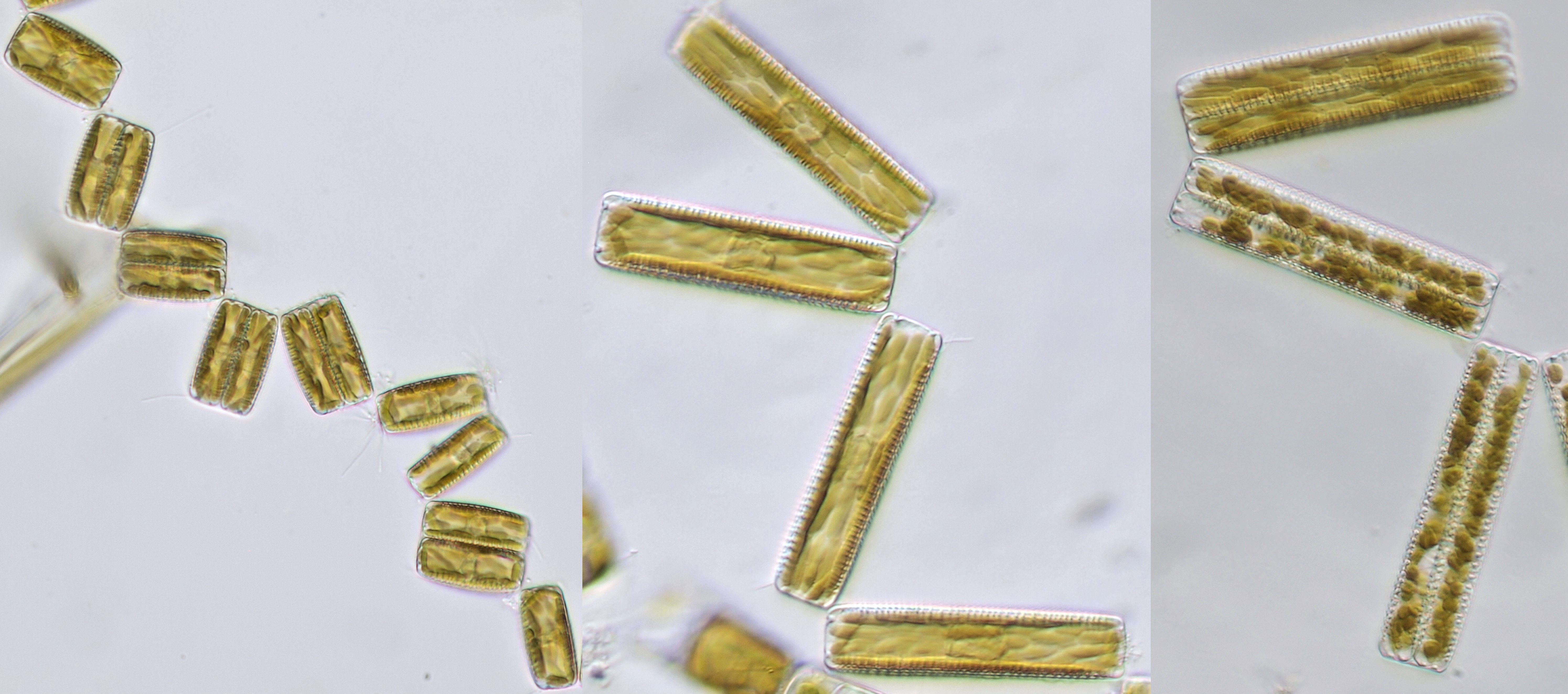
Diatoma from various samples taken from the Maas near Kessel (Limburg, Netherlands). The pictures were made with Zeiss-Winkel Stativ B and Zeiss-Winkel 40/0.65 objective.
Amphora
The term 'amphora' on the internet mainly refers to ancient Greek jars and vases. With a little imagination, the diatom Amphora looks a bit like a vase. Amphora has an oval shape with flattened ends and the size varies from 5 - 100 μm.
Amphora. Objective: Leitz Fluotar 40/0.70.
Amphora. Left and center specimen photographed with Zeiss-Winkel objective 40/0.65. The left specimen was from a sample taken from the Maas near Kessel. Right from a sample taken from the Neckar River near Heidelberg, Olympus objective 40/0.65.
Cocconeis
Cocconeis is a oval shaped diatom that lives epiphytically on filamentous algae.
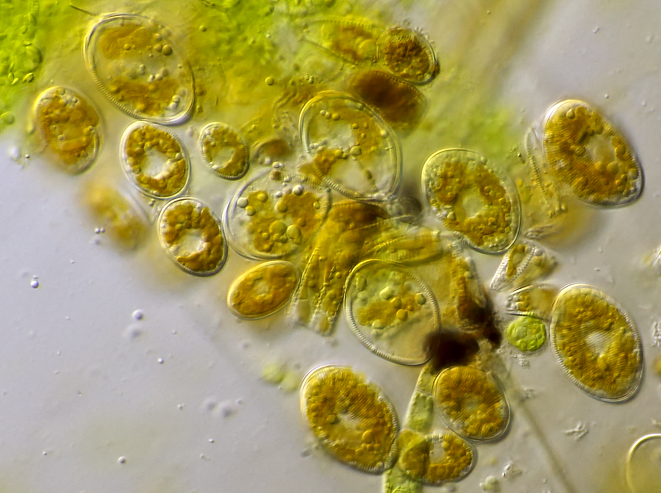
Cocconeis, photographed with Carl Zeiss Apo 40/1.0.
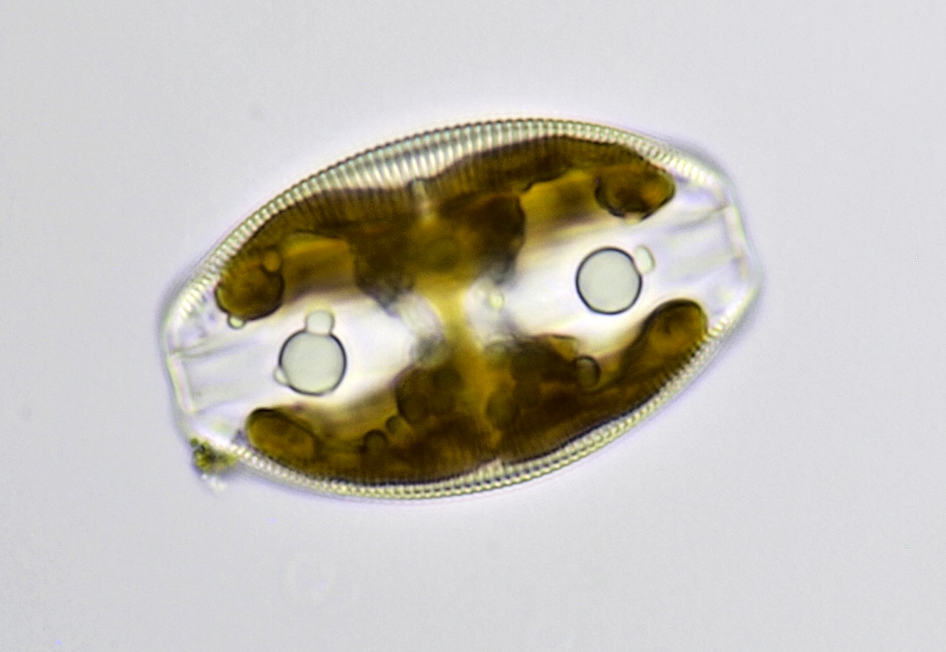
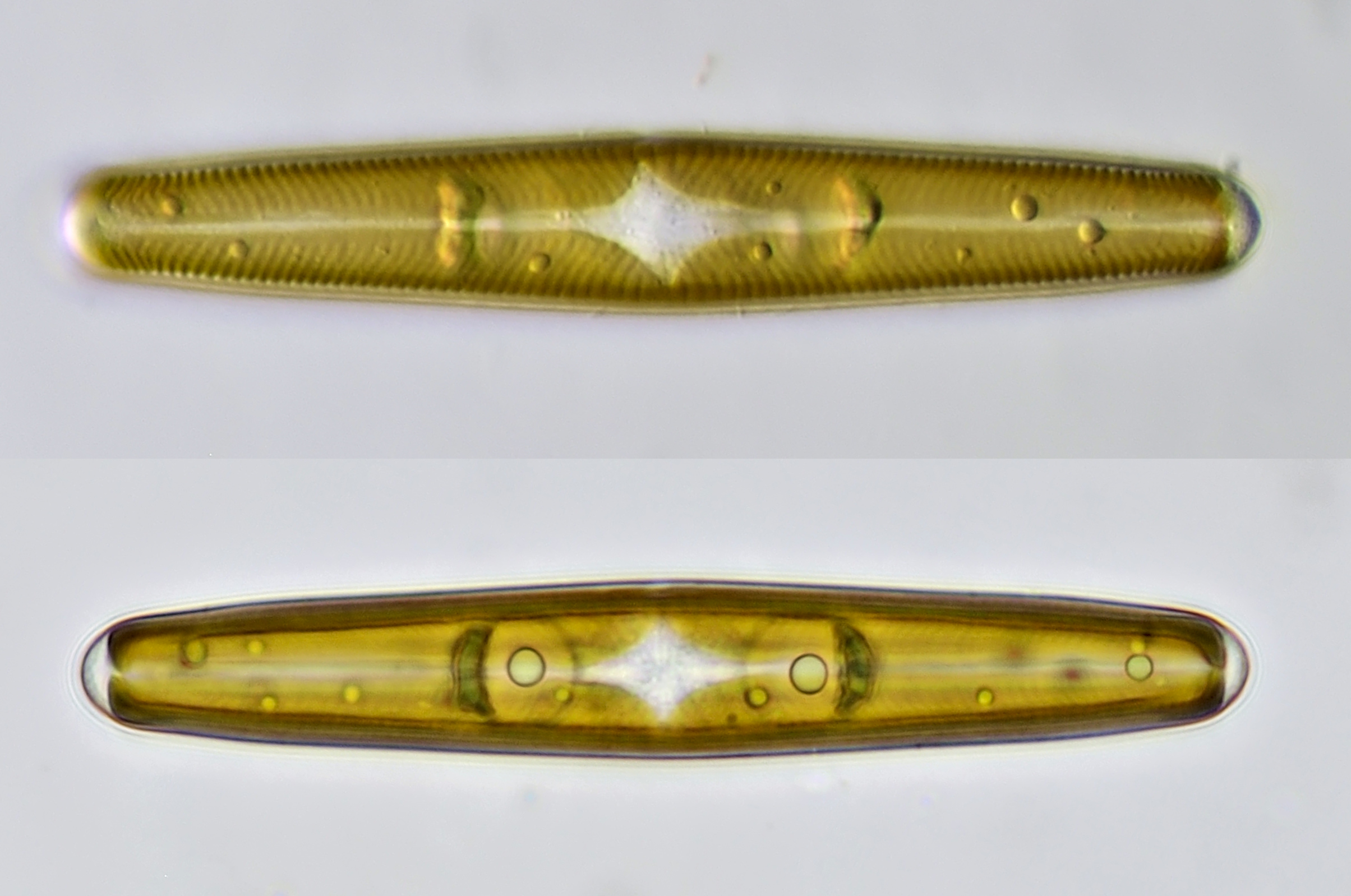
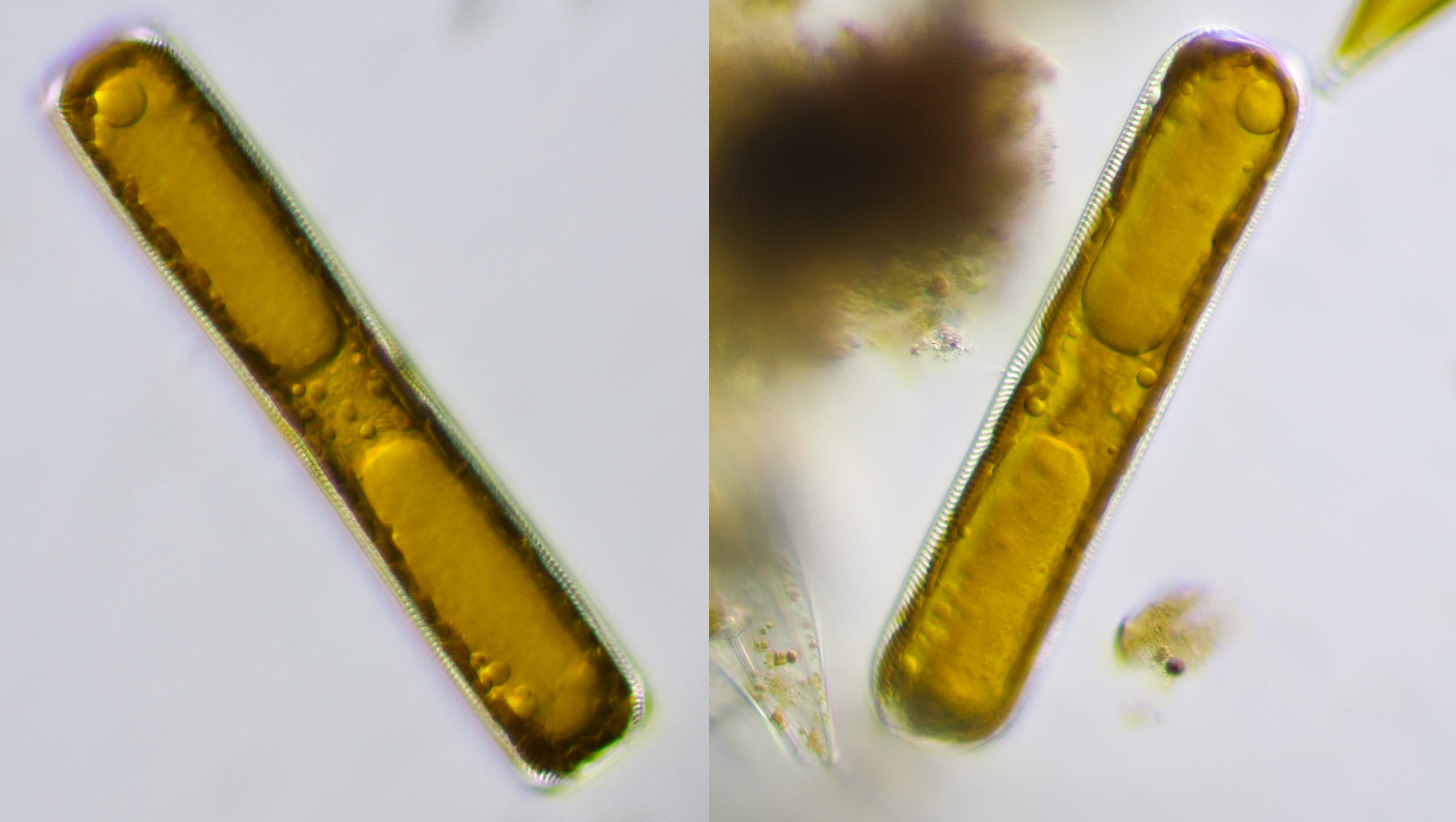
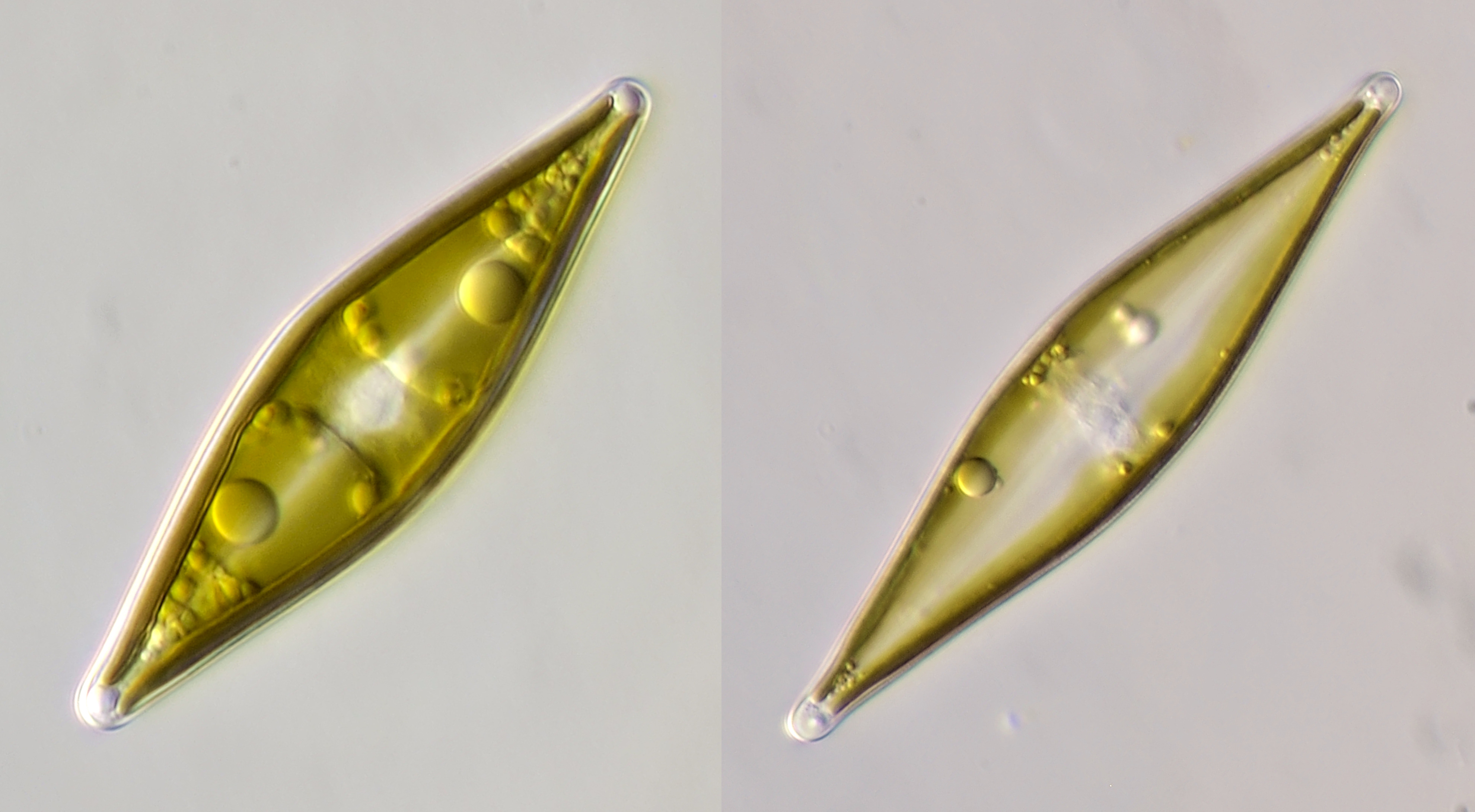
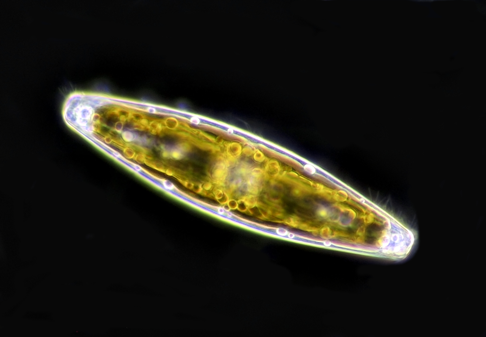
Pinnularia
A pennate diatom that varies in size from 20 - 300 μm and is most widely distributed.
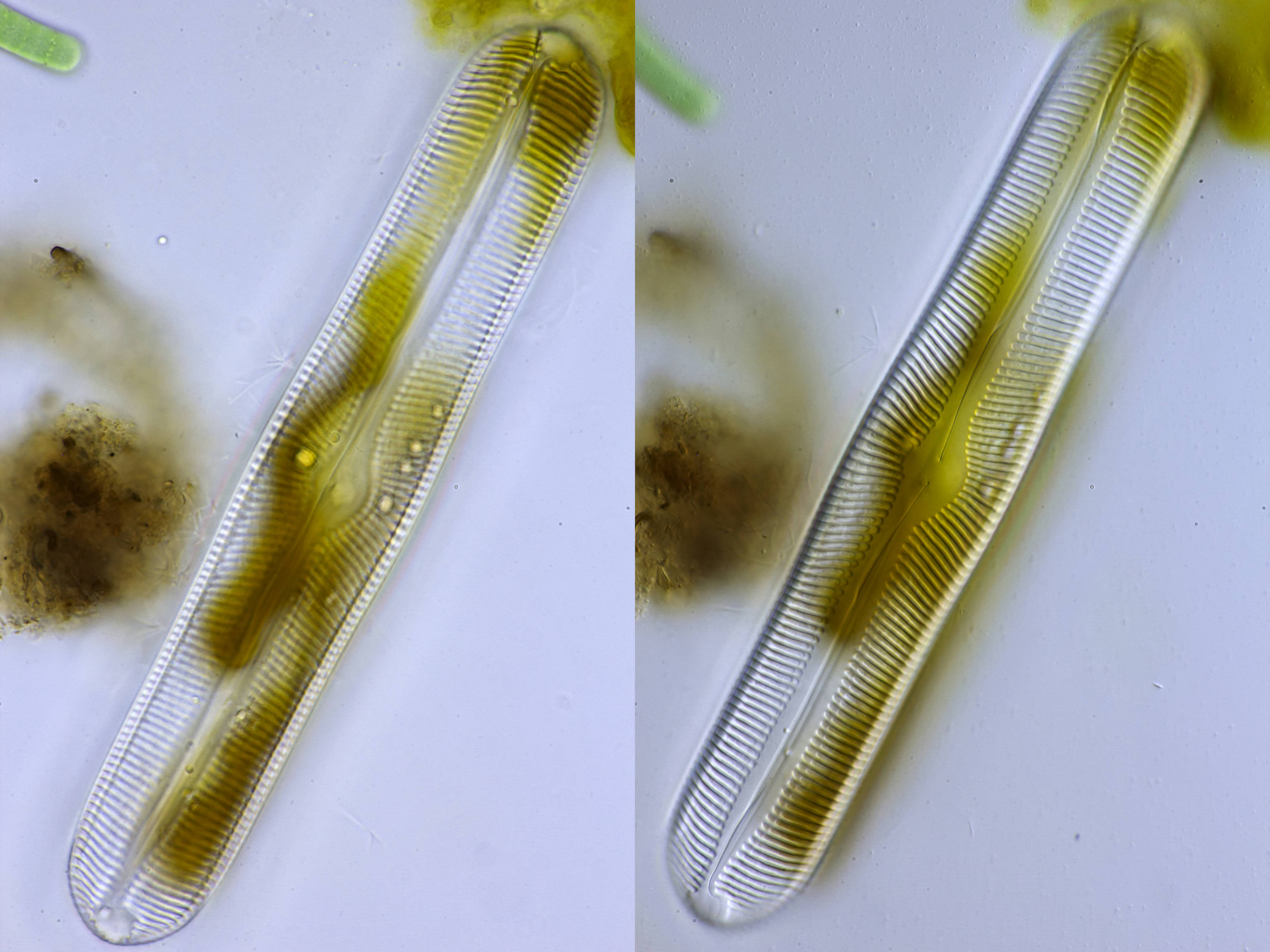
Pinnularia, photographed in normal brightfield (left) and oblique illumination (right). Objective: Carl Zeiss Apo 40/1.0.
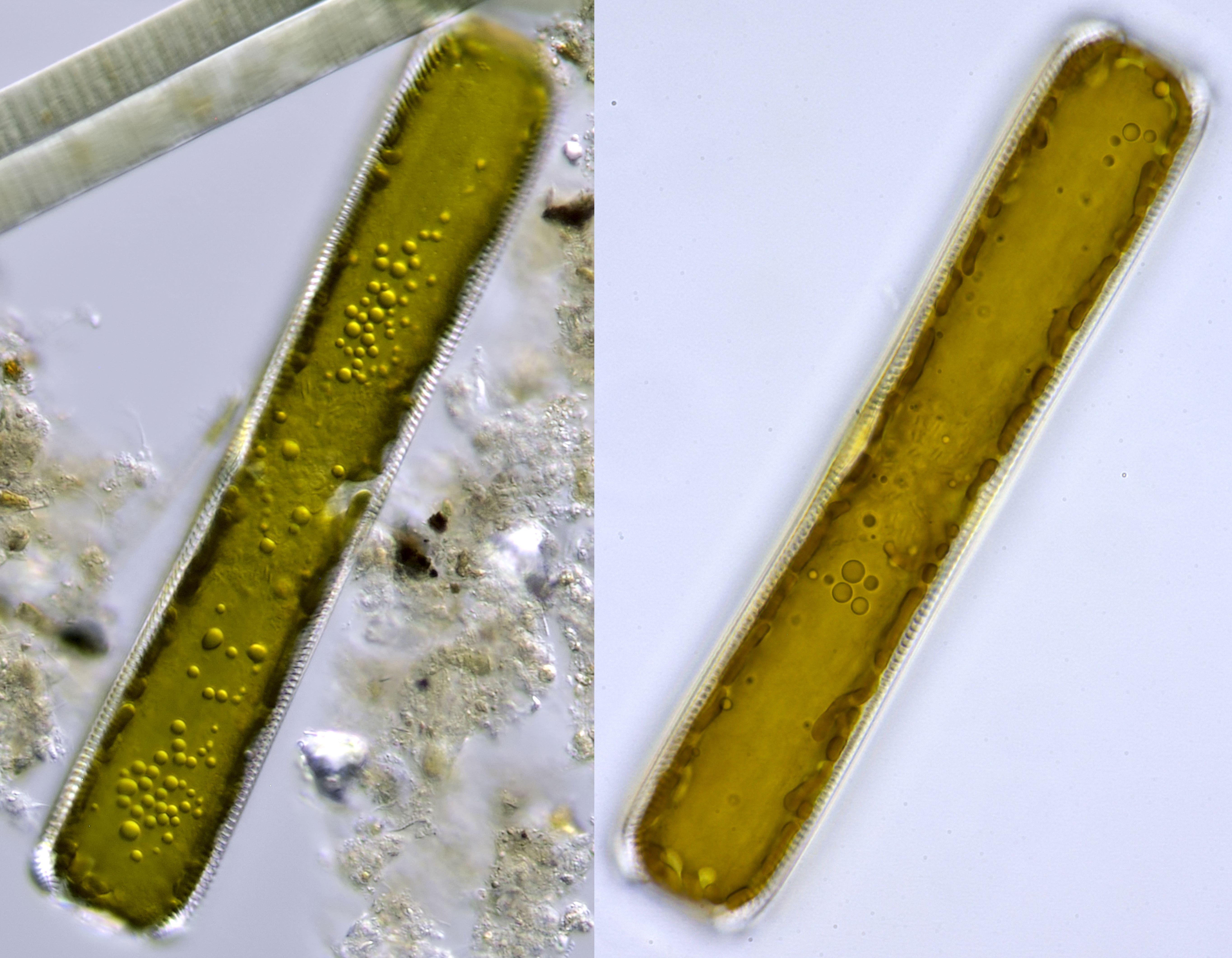
Pinnularia. Left: photographed with oblique illumination and Leitz Fluotar 40/0.70. Oil droplets are clearly visible. Right: brightfield image from another specimen fotographed with Carl Zeiss Apo 40/1.0.
Pinnularia, the same specimen photographed in oblique illumination (upper image) and brightfield illumination (lower image). Objective: Zeiss-Winkel 40/0.65.
Pinnularia, photographed in oblique illumination. Objective: Zeiss-Winkel 40/0.65.
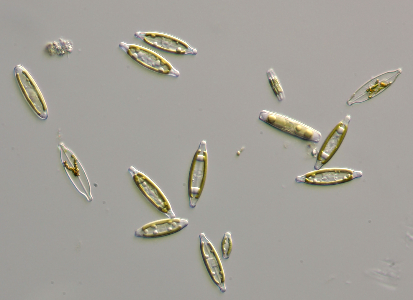
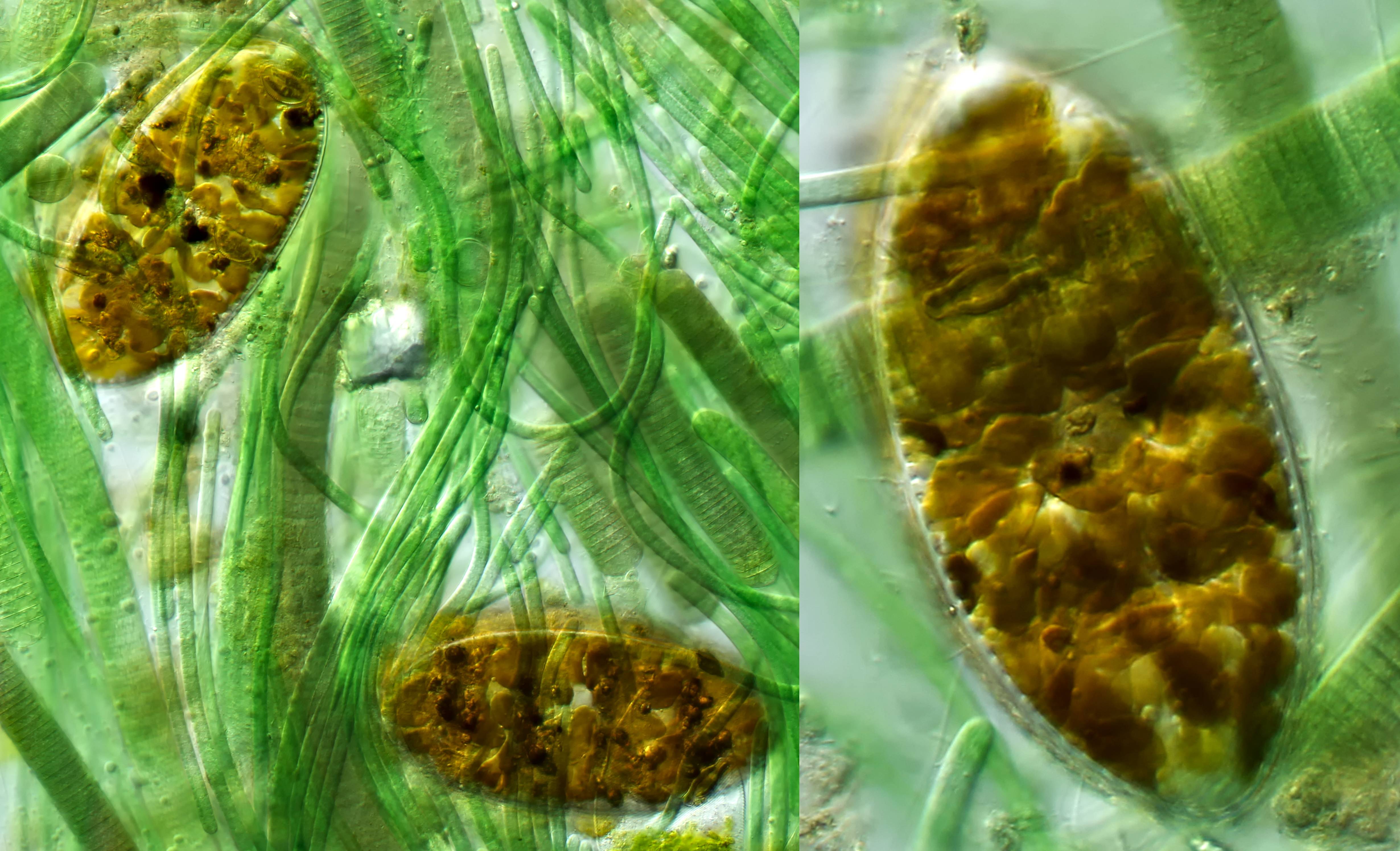
Navicula
It's fascinating to see Navicula move throught micro-space like little boats. Navicula means 'little ship' in Latin.
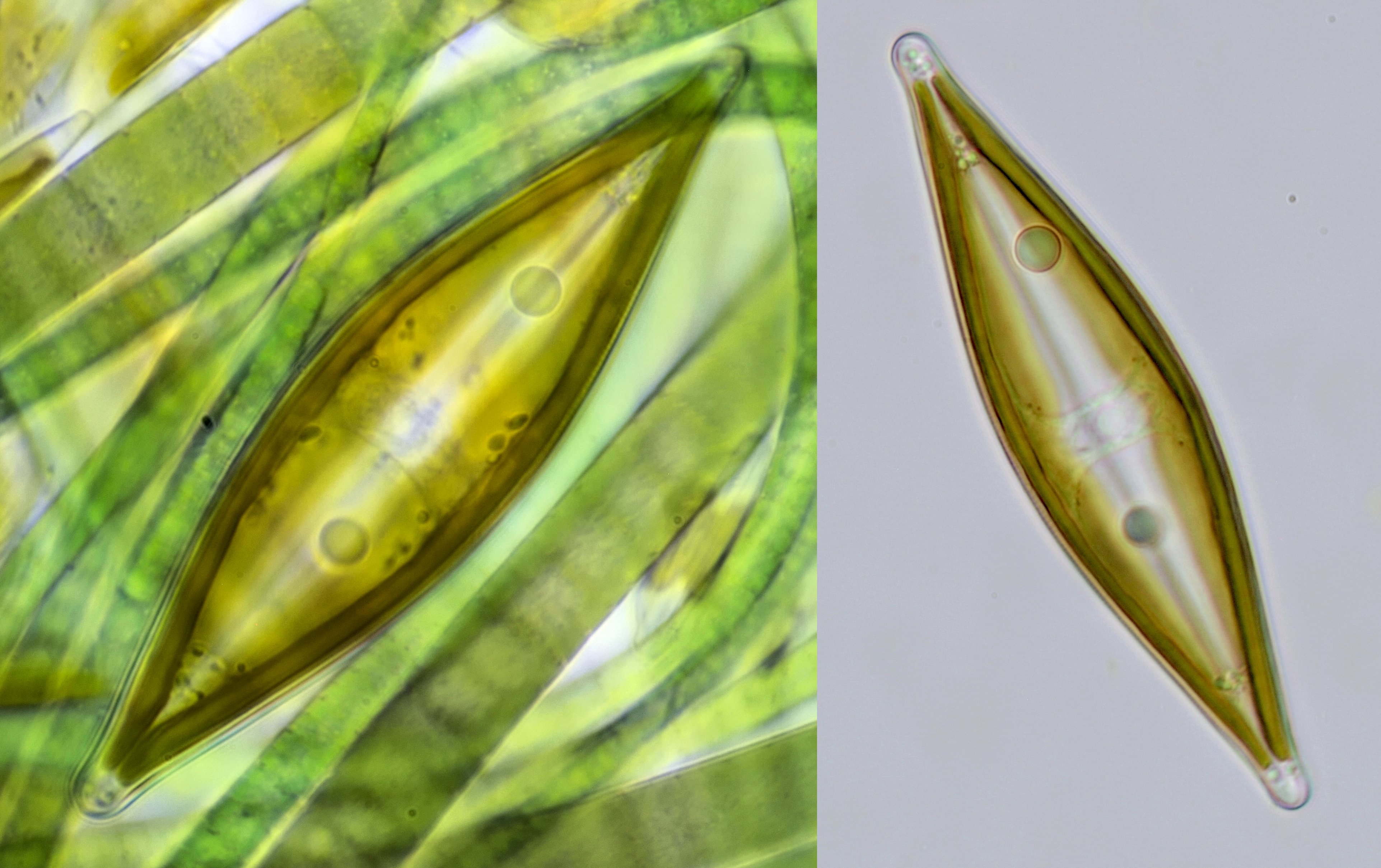
Navicula. Left, hiding between cyanobacteria, objective Zeiss Neofluar 40/0.75. Right: objective Zeiss-Winkel 40/0.65.
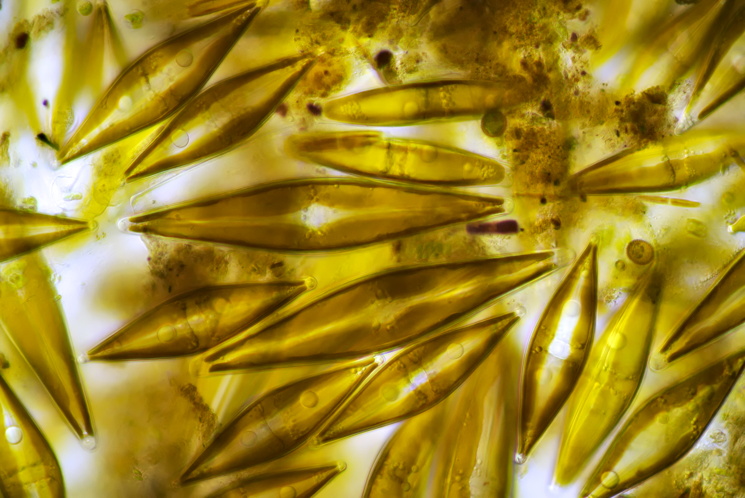
Clustering of Navicula. Objective: Zeiss-Winkel 40/0.65. Camera: Nikon 1 J1.
Video of Navicula on the move. Objective: Zeiss-Winkel 40/0.65.
Navicula from a dune lake called 't Wed located in the Kennemer Dunes. Objective: Leitz Fluotar 25/0.55.
Navicula photographed with oblique illumination. Objective: Zeiss-Winkel 40/0.65.
Navicula, photographed in darkfield illumination. Objective: Leitz 25/0.50.
Fragilaria
Fragilaria, a colony-forming diatom in which the individual cells form filaments.
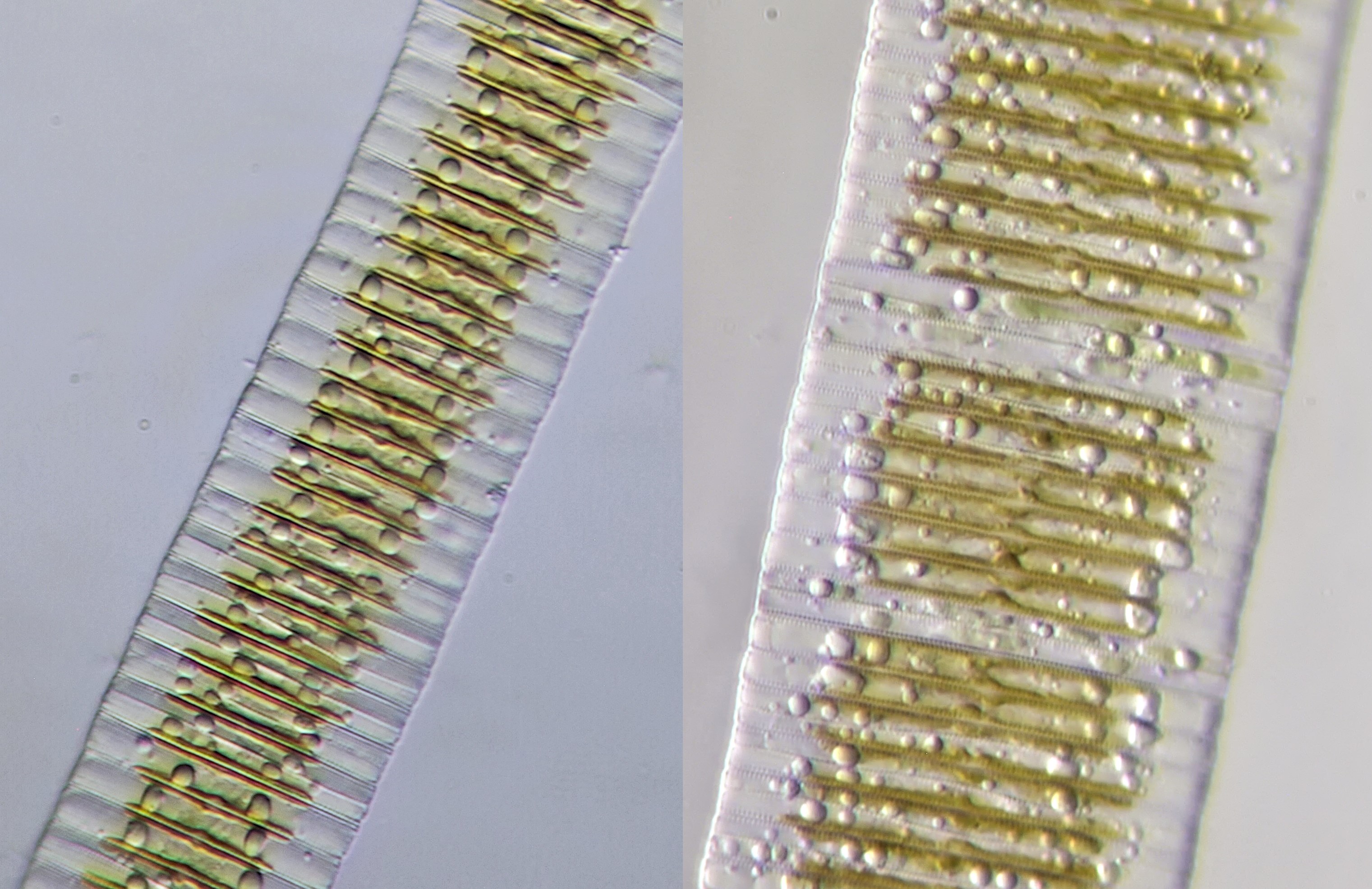
Fragilaria, photographed with Zeiss-Winkel 40/0.65.
Fragilaria, left photographed with Zeiss-Winkel 40/0.65 and oblique illumination. Right in phasecontast with Leitz EF 40/0.65 Phako.
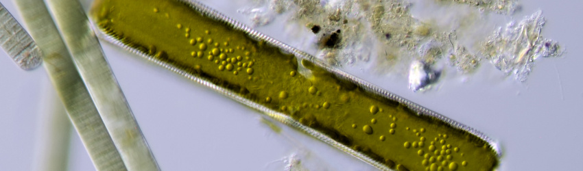
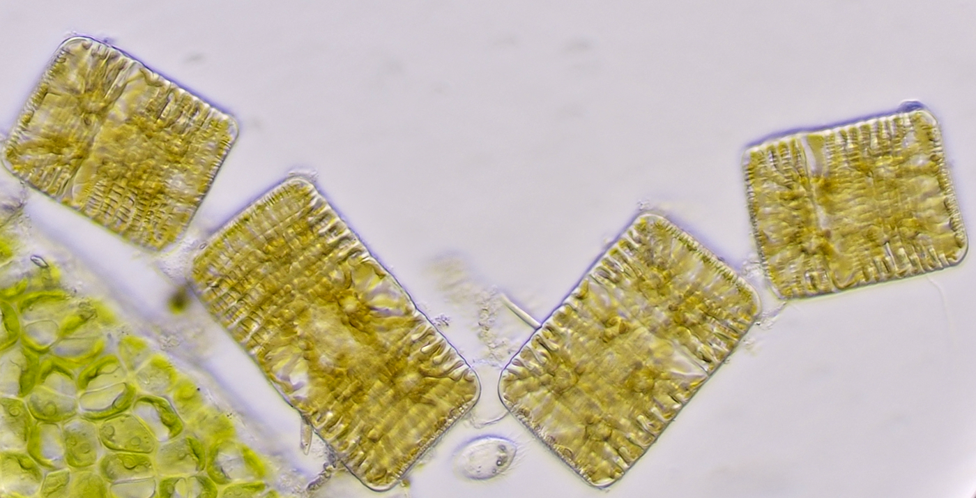
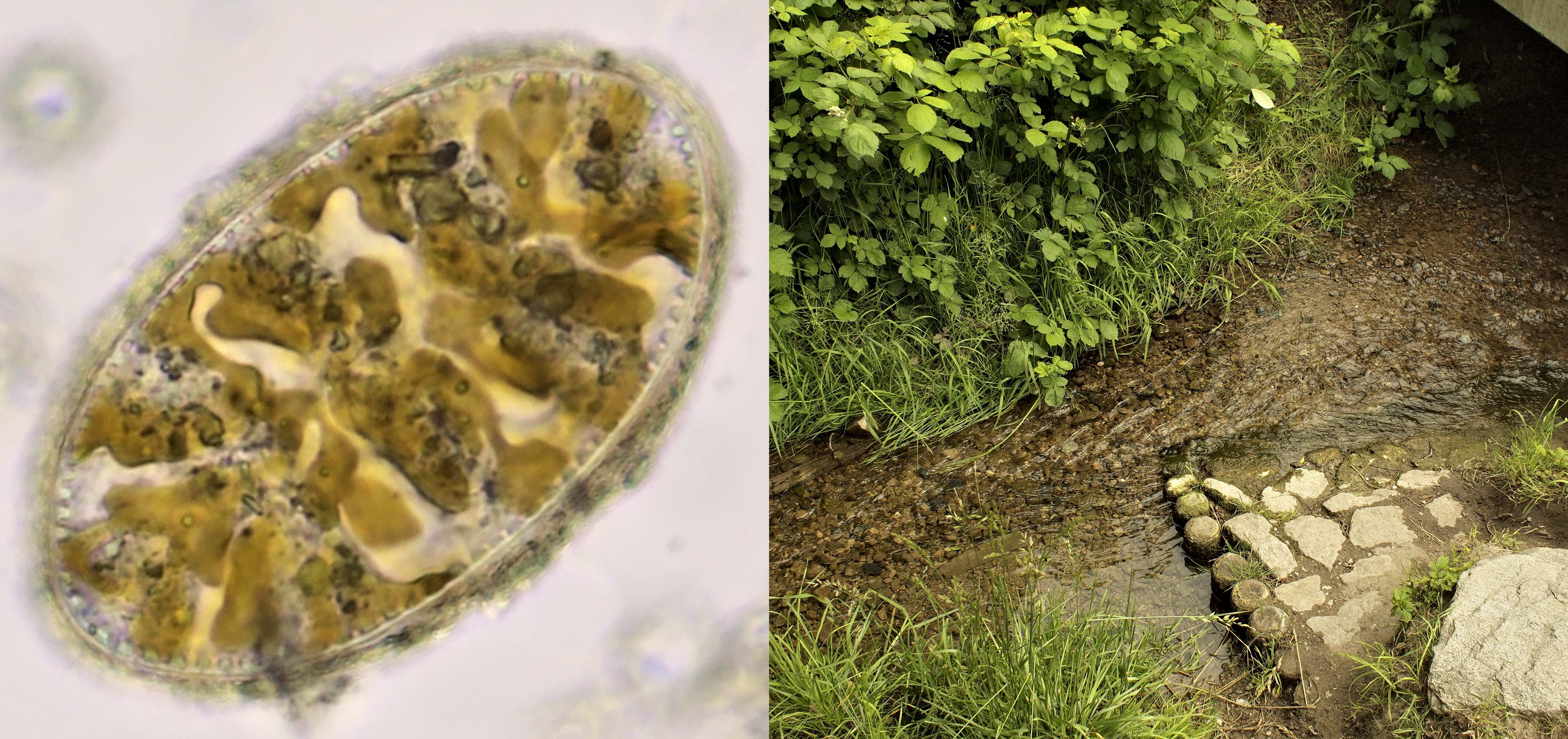
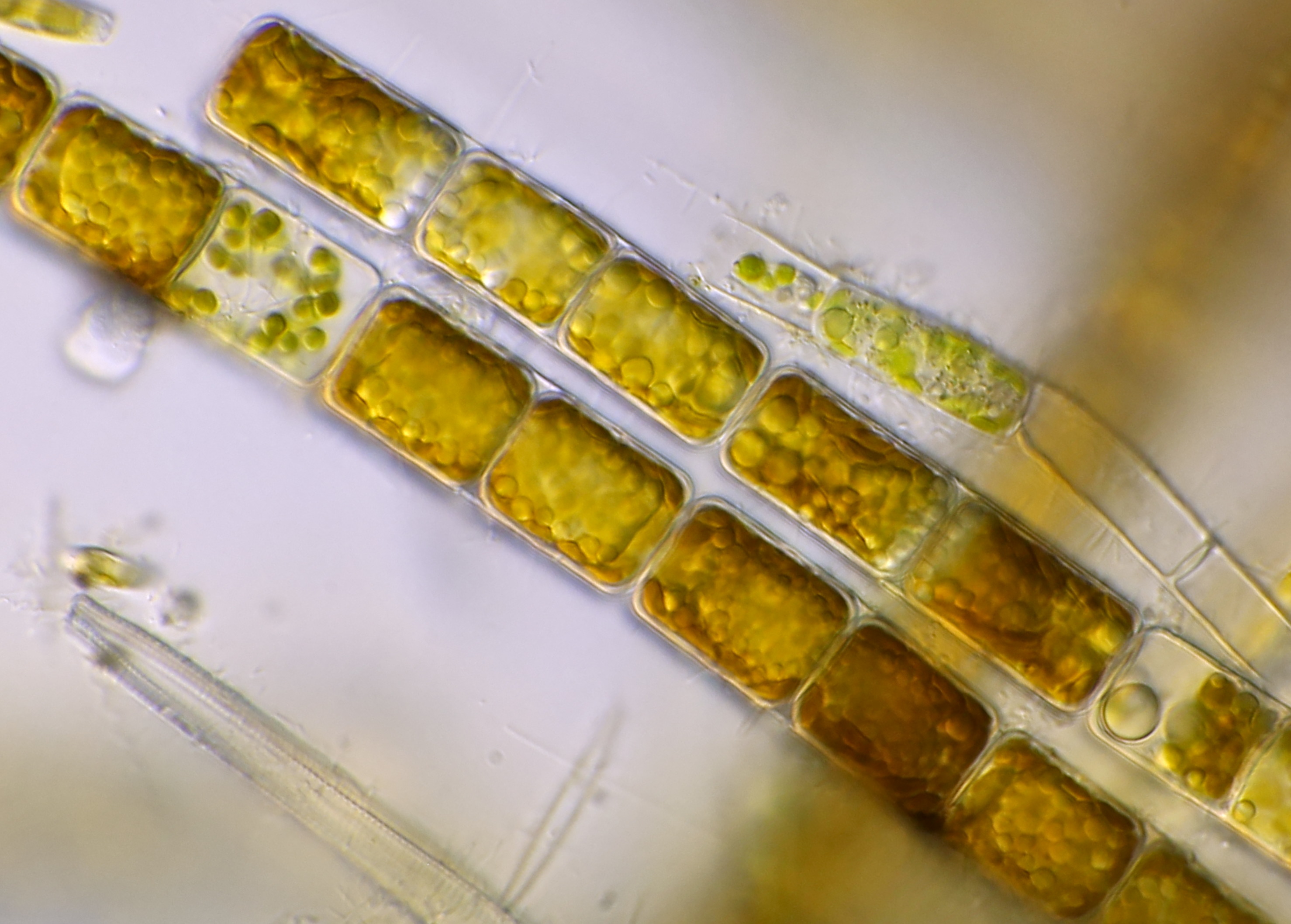
Pleurosira
Pleurosira, a diatom that forms chains of very large cells.
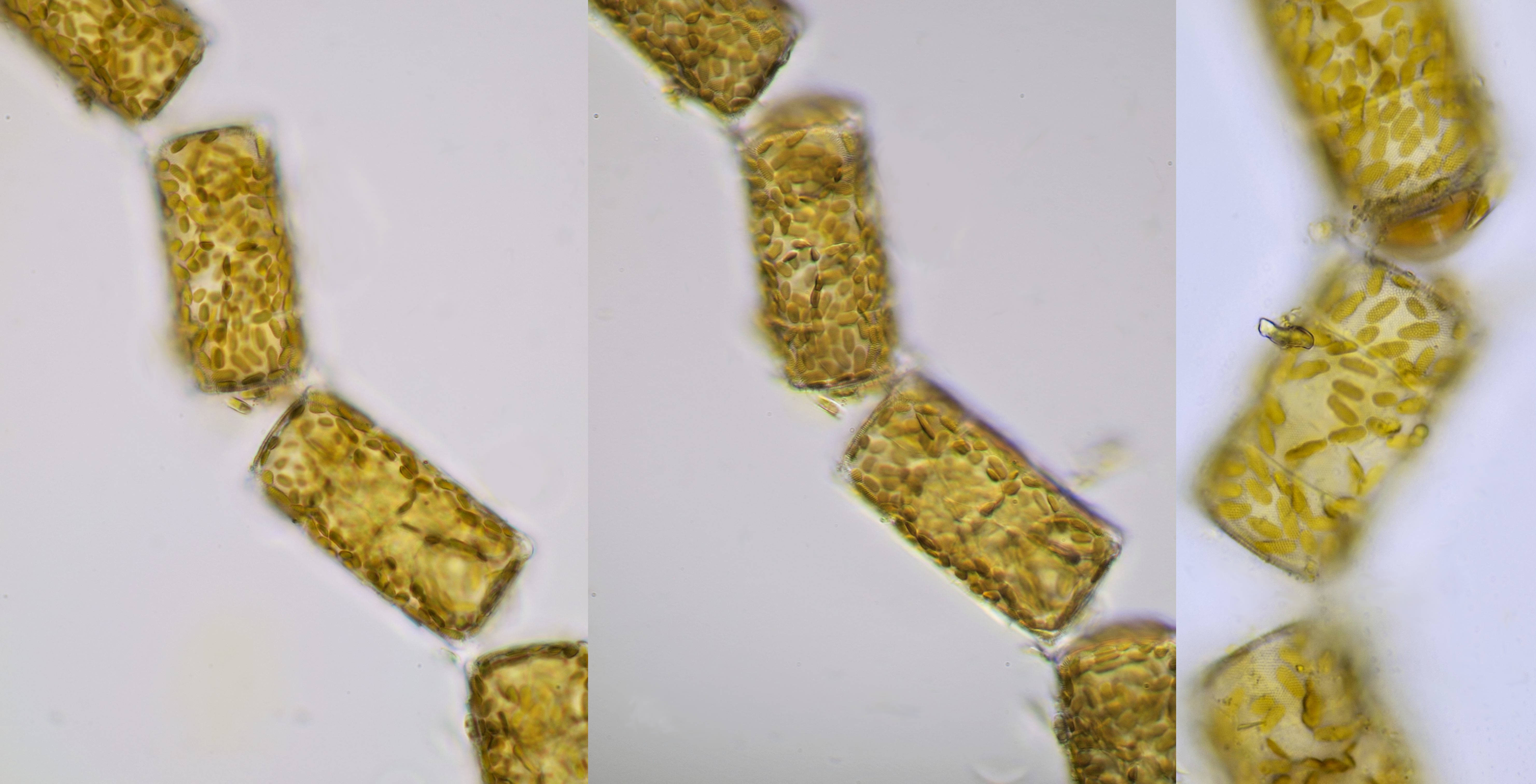
Pleurosira. The sample was taken from the Maas at the location 'de loswal' in Kessel (Limburg, Netherlands). Left and middle photo: Zeiss-Winkel 40/0.65. Photo on the far right: Carl Zeiss Neofluar 40/0.75.
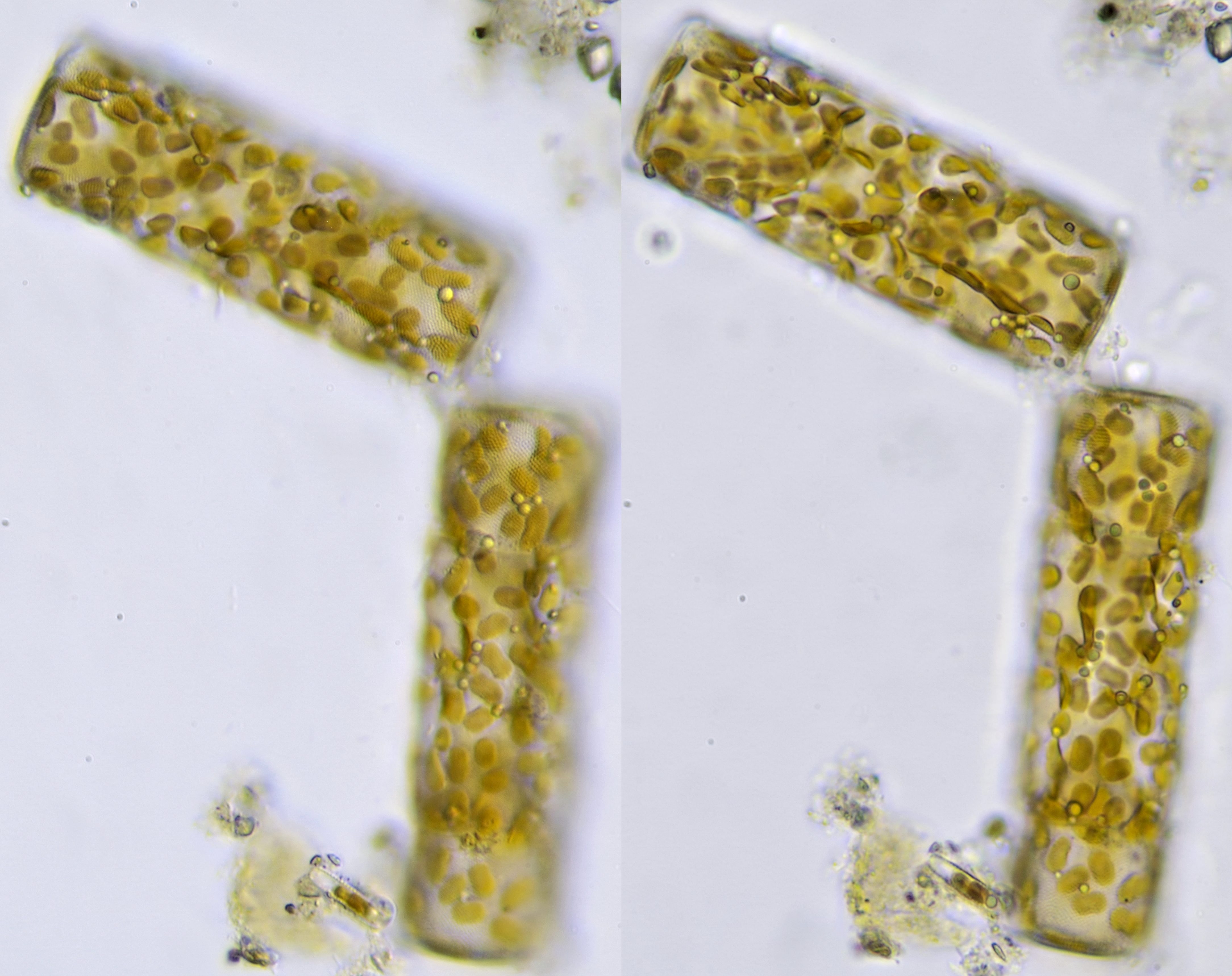
Pleurosira from a sample taken from the Neckar River near Heidelberg. Photographed with Olympus objective 40/0.65 and focused on different focal planes.
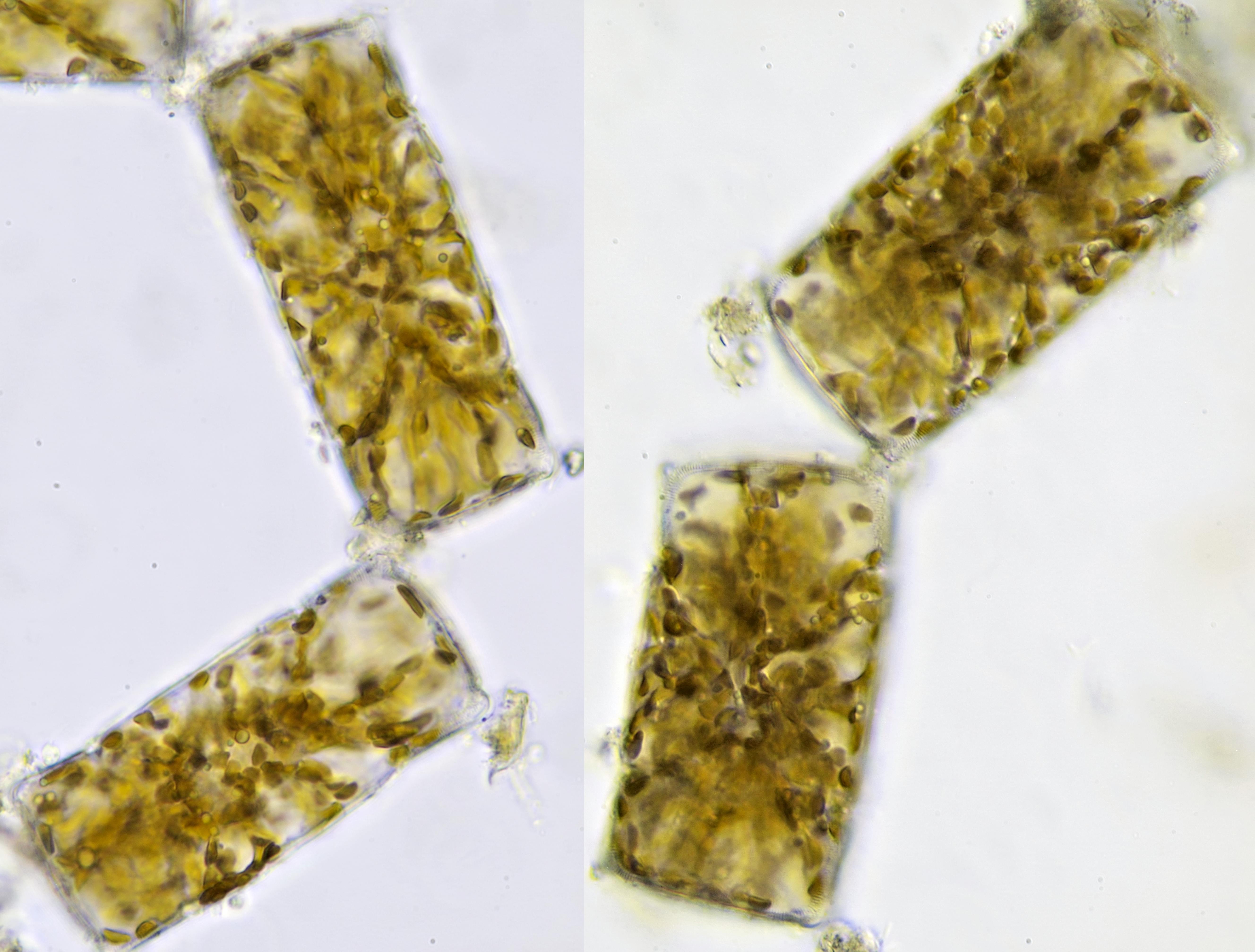
Pleurosira from a sample taken from the Neckar River near Heidelberg. The photos were taken during a holiday in Heidelberg with the Olympus HSA microscope that I had taken with me as a travel microscope. Objective: Olympus 40/0.65.
Pleurosira from the North Sea near the province of Zeeland. Objective: Olympus 40/0.65.
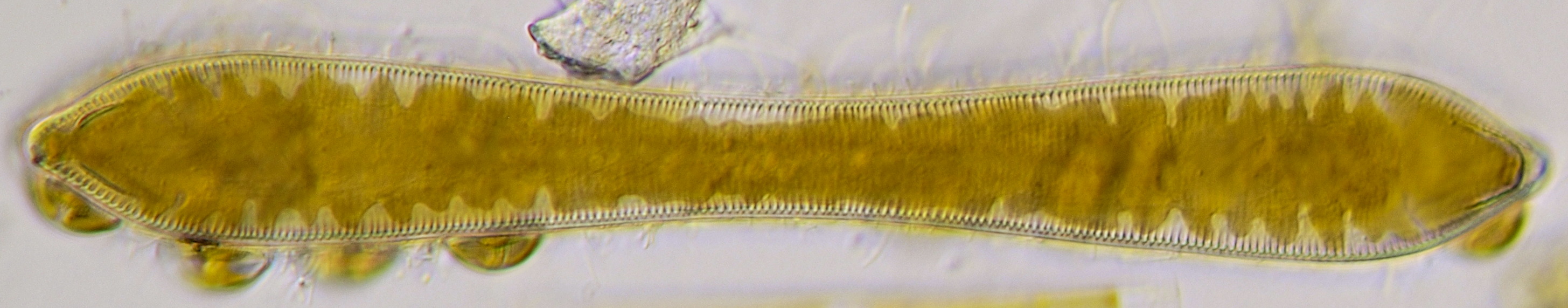
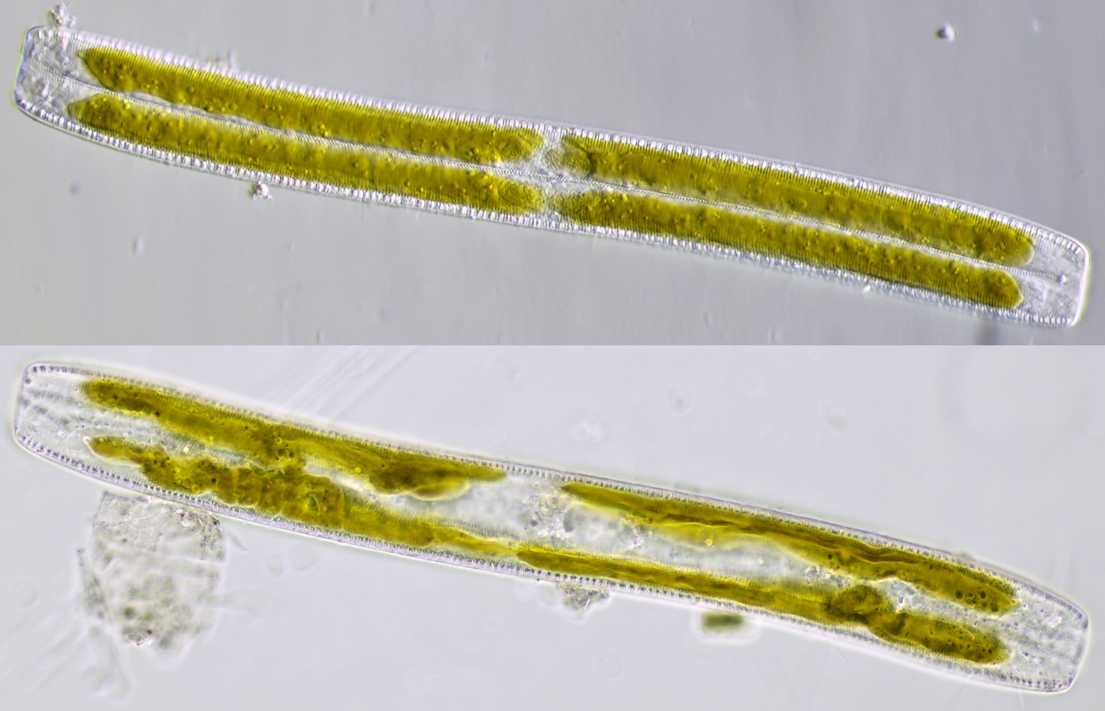
Cymatopleura
This diatom has an elliptical shape with often a narrowing in the middle.
Cymatopleura, photographed with Euromex 40/0.65.
Cymatopleura from a ditch in Zevenaar. Photographed with Carl Zeiss Apo 40/1.0.
Cymatopleura elliptica. Objective: Leitz Pl Apo 25/0.65.
Cymatopleura elliptica from the Gembdenbach, a small stream near Jena. Photographed during holidays with the Olympus HSA and objective 40/0.65.
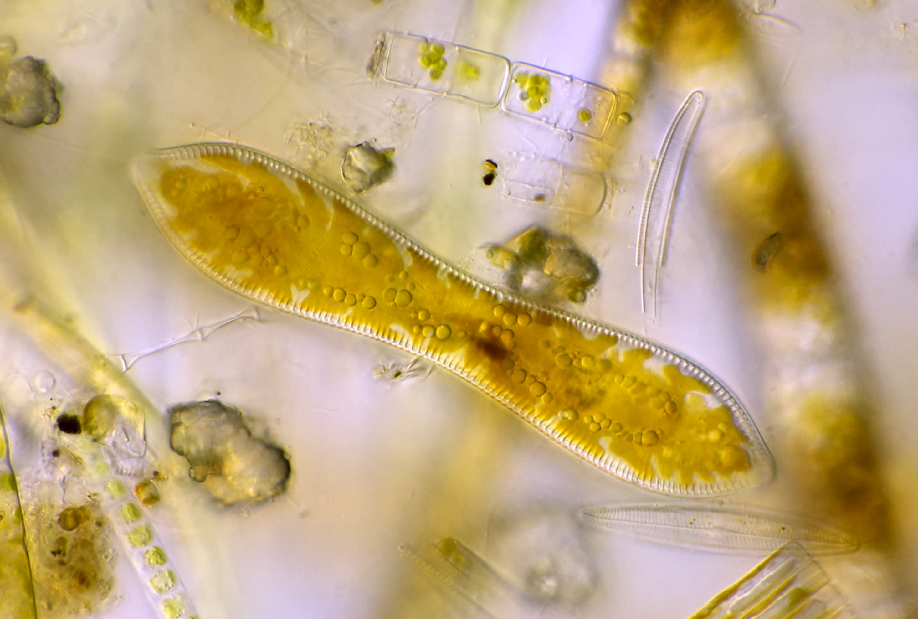
Cylindrotheca closterium
A marine diatom with a special shape.
Cylindrotheca closterium photographed in phasecontrast with Carl Zeiss Neofluar Ph2 40/0.75.
Nitzschia
Very elongated and narrow shaped diatoms that vary in lenght from 30 - 500 μm. Many species produce toxic substances.
Two different specimens of Nitzschia photographed with Leitz Fluotar 25/0.55 in oblique illumination (top) and with Leitz Pl Apo 25/0.65 in annular illumination (bottom).
Melosira
A filamentous diatom with radial symmetry that tolerates poor water quality.

Melosira. Two different filaments photographed with Carl Zeiss 63/0.80 (upper image) and Zeiss-Winkel 40/0.65 (lower image).
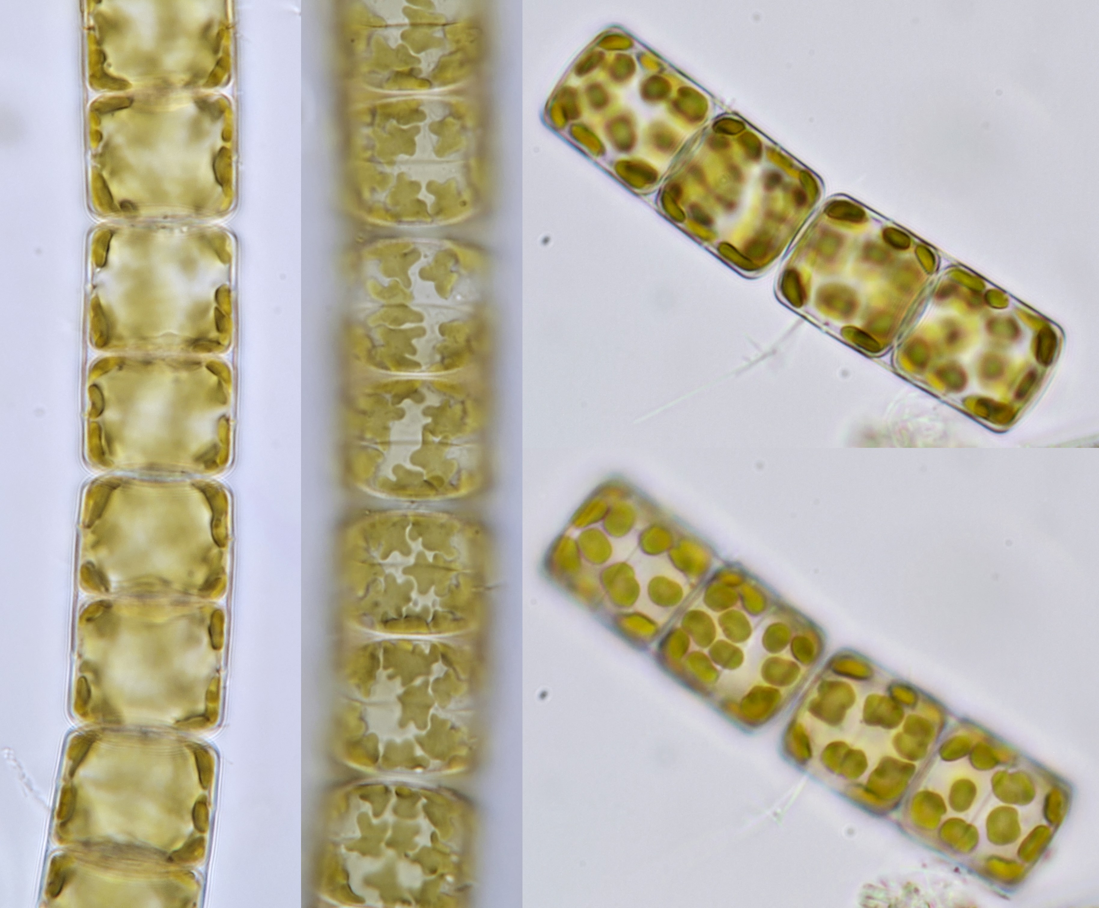
Melosira from the Maas near Kessel (Limburg, Netherlands). Two filaments focused at different depths. Left photos: Carl Zeiss Apo 40/1.0. Right photos: Zeiss-Winkel 40/0.65.
Melosira from a ditch in Zevenaar photographed with Carl Zeiss Apo 40/1.0.
Cyclotella
A centric diatom with a diameter of 4 - 40 μm.
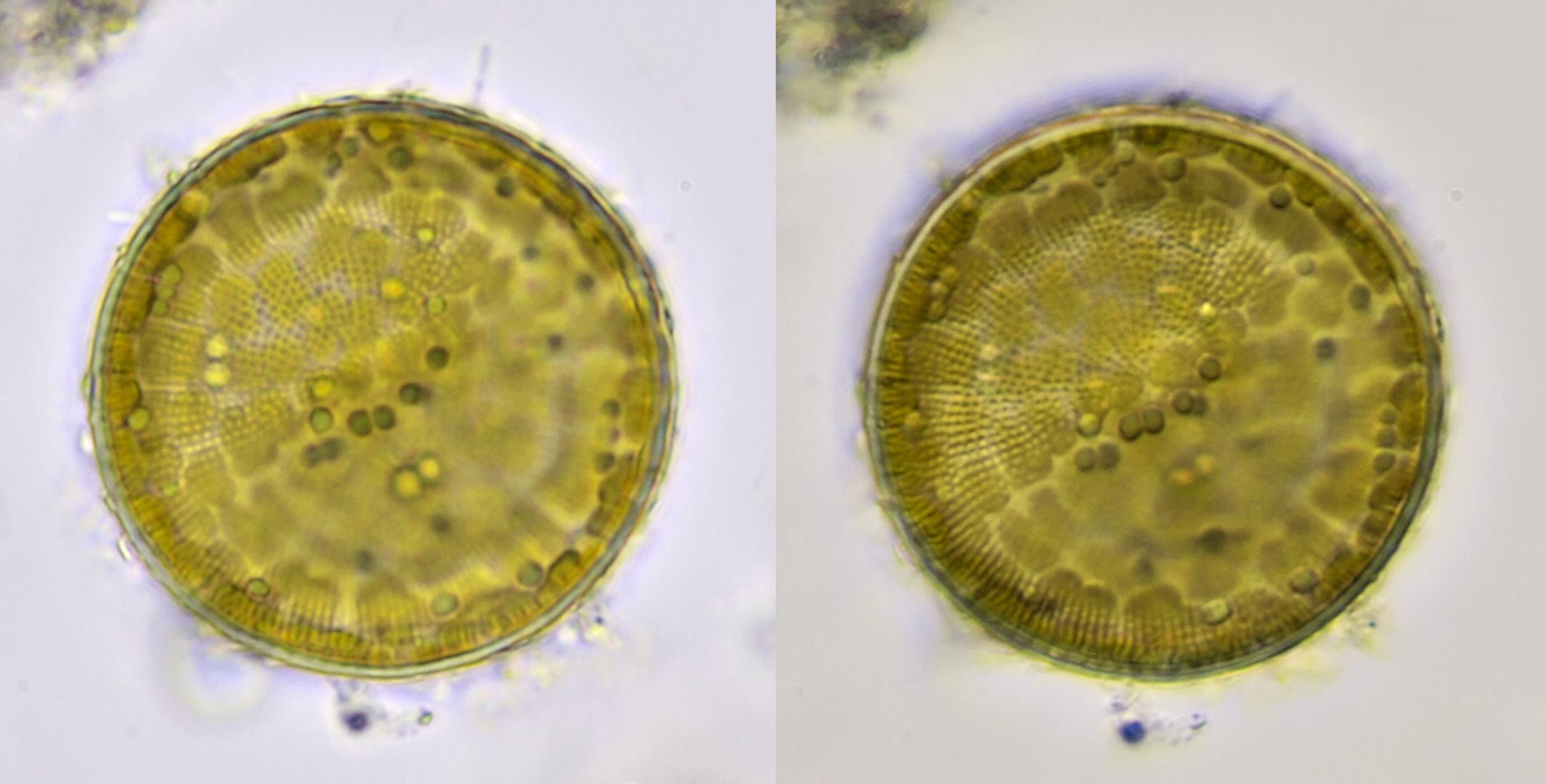
Cyclotella from the Neckar near Heidelberg photographed in brightfield (left) and oblique illumination (right). Objective: Olympus 40/0.65.
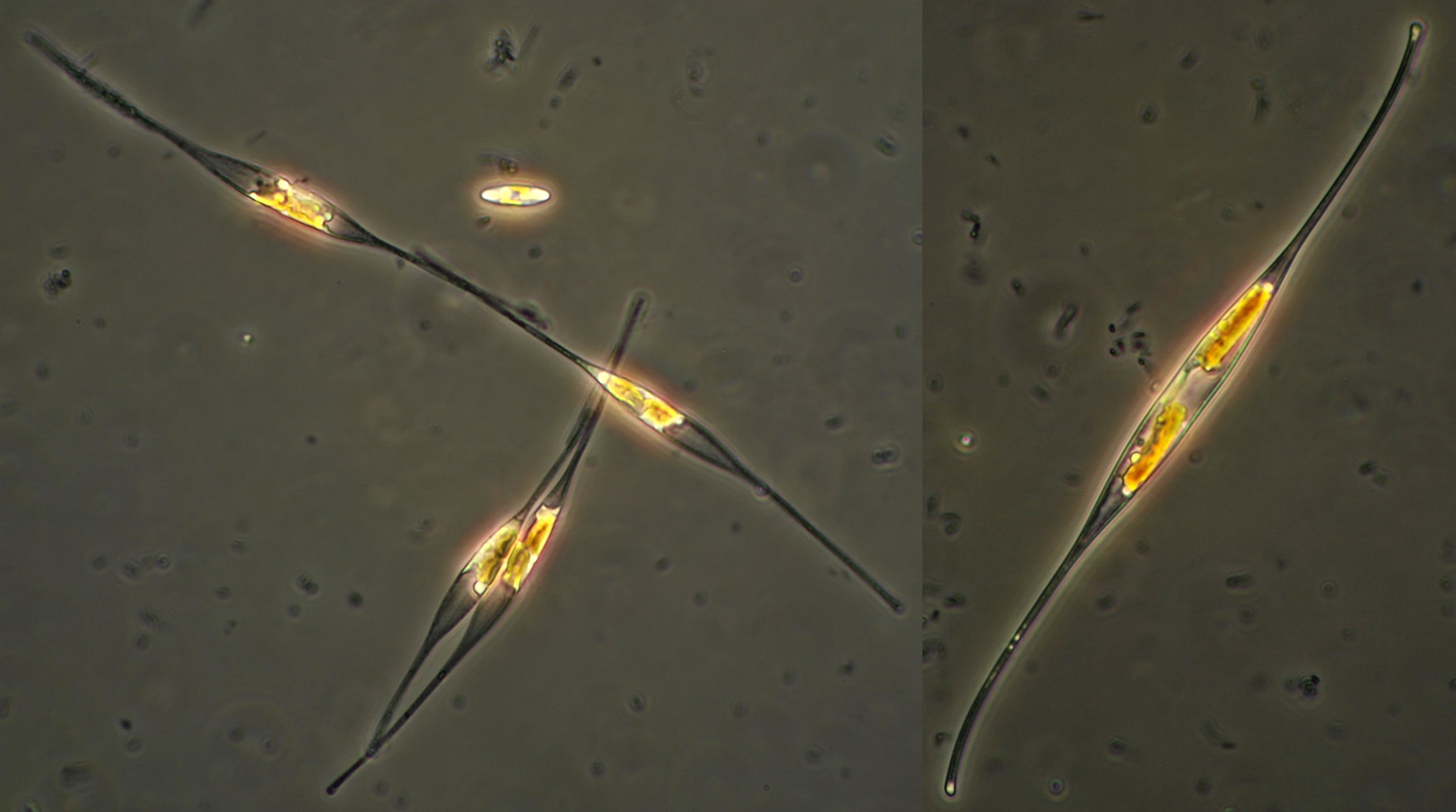
Gyrosigma
A diatom with a sigmoid shape, ranging in size from 60 - 180 μm.
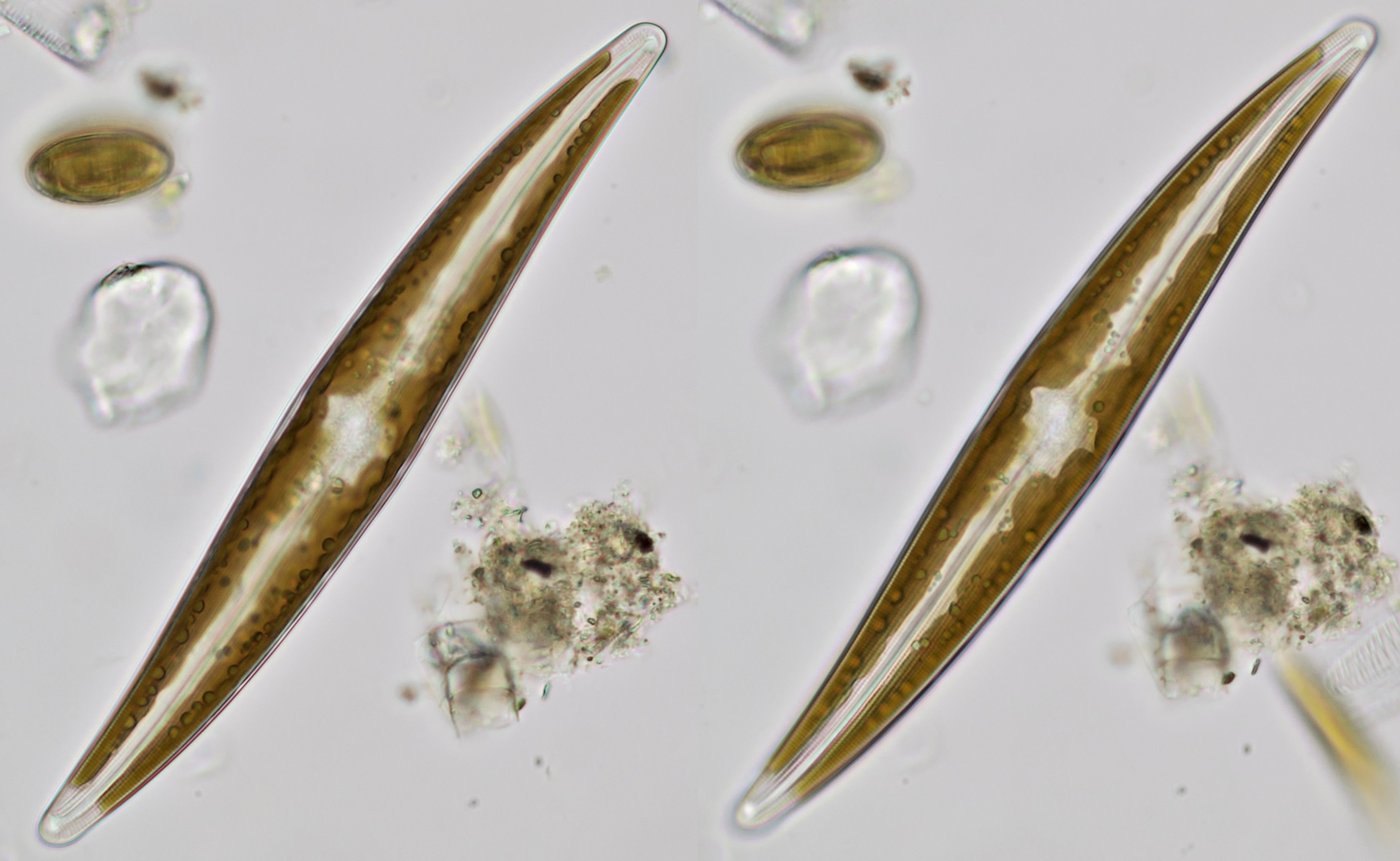
Gyrosigma from a sample taken from the Maas near Kessel. The same specimen was photographed twice with a small difference in focus. Objective: Zeiss-Winkel 40/0.65.
Gomphonema
A diatom ranging in size from 10 -130 μm which is often attached to other algae by a stem.
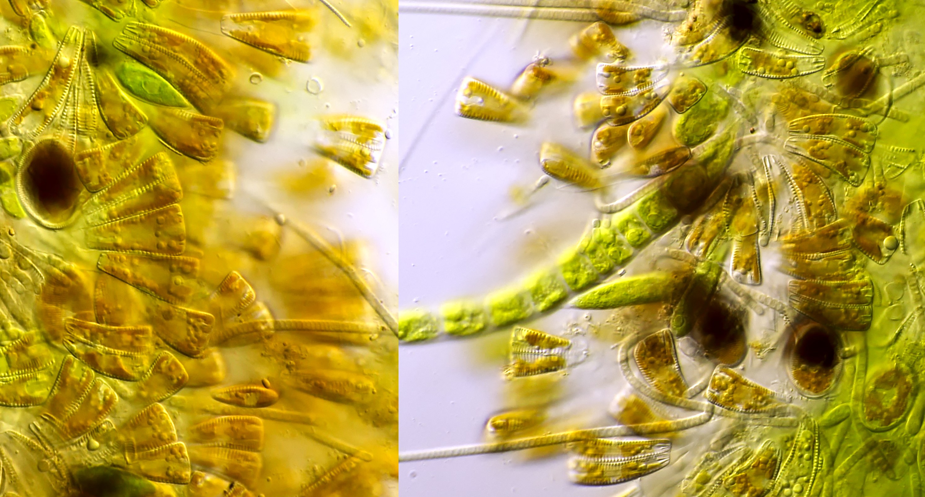
Gomphonema from river Amstel. Objective: Carl Zeiss Apo 40/1.0.
Literature
Linne von Berg, KH., Hoef-Emden, K., Marin, B., Melkonian, M. (2012). Der Kosmos Algenführer. Stuttgart: Franckh-Kosmos-Verlags-GmbH & Co.