It is not that often that I look at blood under the microscope. It usually happens when I get injured and have the opportunity to get a microscope. After an unfortunate shave, I act quickly so that I can prepare a slide from the blood before it dries up.
If blood is examined microscopically in the laboratory, this is done by means of differential staining. Here, the different types of leucocytes (white blood cells) are stained differently and a discrimination can be made on the basis of cell-specific characteristics. To perform a differential staining, a thin blood smear is made on a clean glass slide and then air dried. This is followed by a fixation step and a staining procedure according to May-Grünwald Giemsa. The next image shows an example of a differential staining.
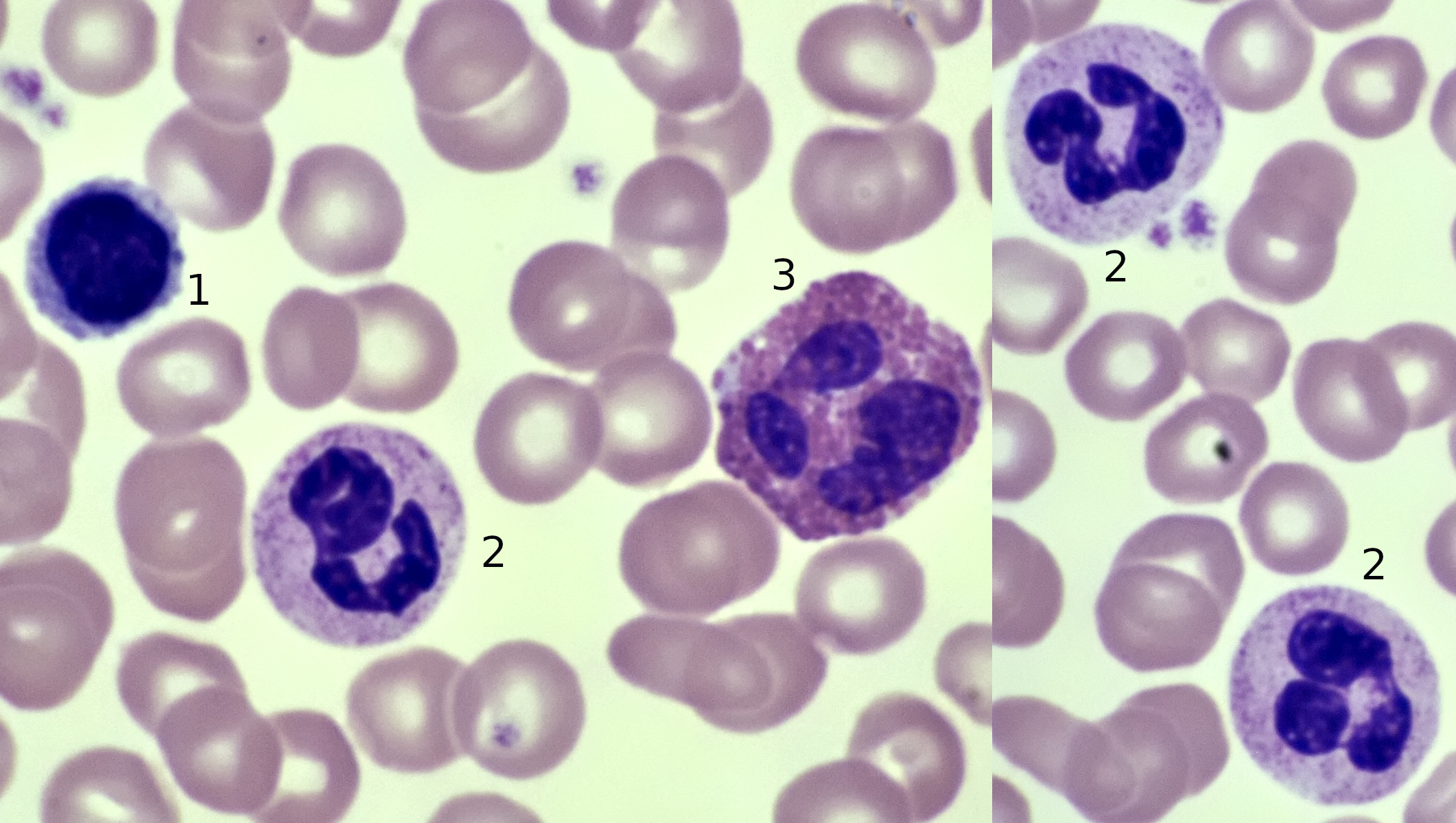
Differential staining of blood showing different types of leucocytes. In the picture, a lymphocyte (1, diameter 8-20 μm), neutrophil granulocytes (2, diameter 11-16 μm) and a eosinophil granulocyte (3, diameter 10-15 μm) can be seen. The erythrocytes (red blood cells) in the background have an average diameter of 7 μm. Objective: Carl Zeiss Apo 40/1.0.
Performing a differential staining of blood is not for the beginner, especially since the interpretation of such a staining should be left to someone with a medical background and knowledge of hematology. As an amateur you can make the different types of leucocytes visible, but that’s it. However, it is also very interesting to examine fresh, unstained blood. Also here, no conclusions should be drawn. When looking at fresh blood under a microscope, detecting leucocytes can be a challenge. Unstained white blood cells are quite difficult to see and only with the help of darkfield or phase contrast microscopy they are easily seen. To make a slide from fresh blood, a small drop of blood is placed on a glass slide and covered with a coverslip. The blood will slowly disperse under the coverslip. It is important to use a very small drop of blood, otherwise the layer of blood will become too thick and many red blood cells will be lying on top of each other.
If you look at the erythrocytes in a slide from fresh blood, you will notice that they have a biconcave shape. They are thinner in the center than at the edge. Due to this form, oxygen is more easily absorbed and released. The shape of the erythrocytes can be clearly in fresh blood, even better than in a stained slide. In many pictures and films, erythrocytes are displayed in a bright red colour. But in reality, the individual red blood cells are not that red and the colour is more like salmon pink. Regarding the leucocytes, some can move actively like an amoeba. It's fascinating to observe, see also the video at the bottom of this page.
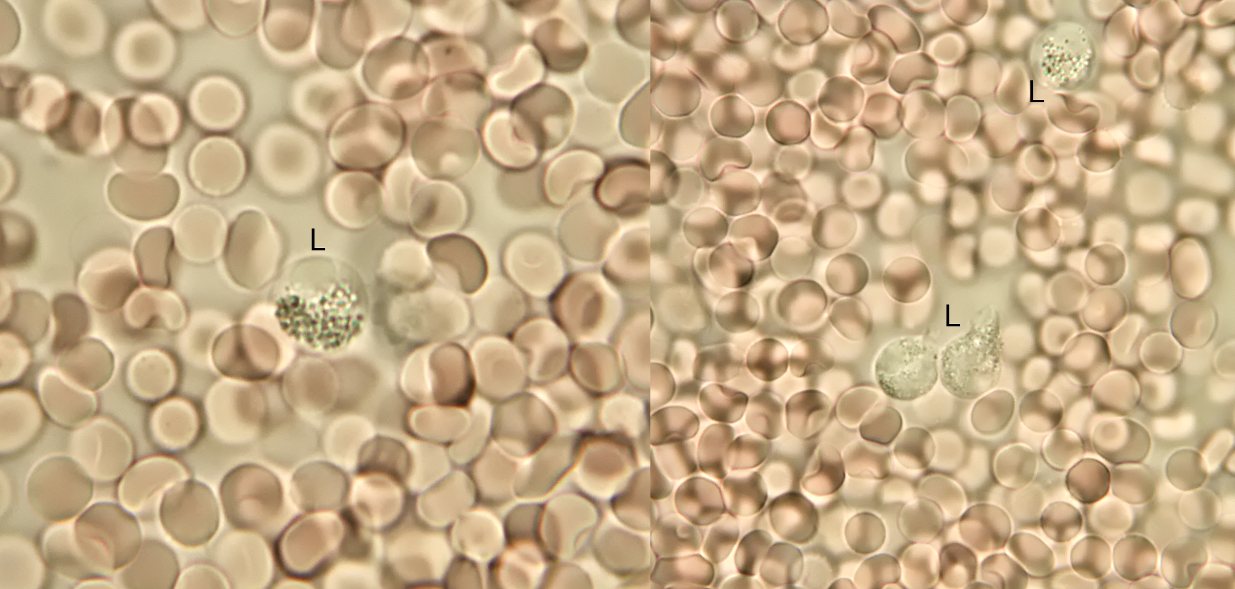
Slide from fresh blood. Note the biconcave shape of some of the erythrocytes. Also, a few leucocytes (L) can be seen. Objective: 100/1.25.

Image of fresh blood taken with circular oblique illumination. The biconcave shape of the erythocytes is clearly visible here. Objective: Carl Zeiss Plan 25/0.45.
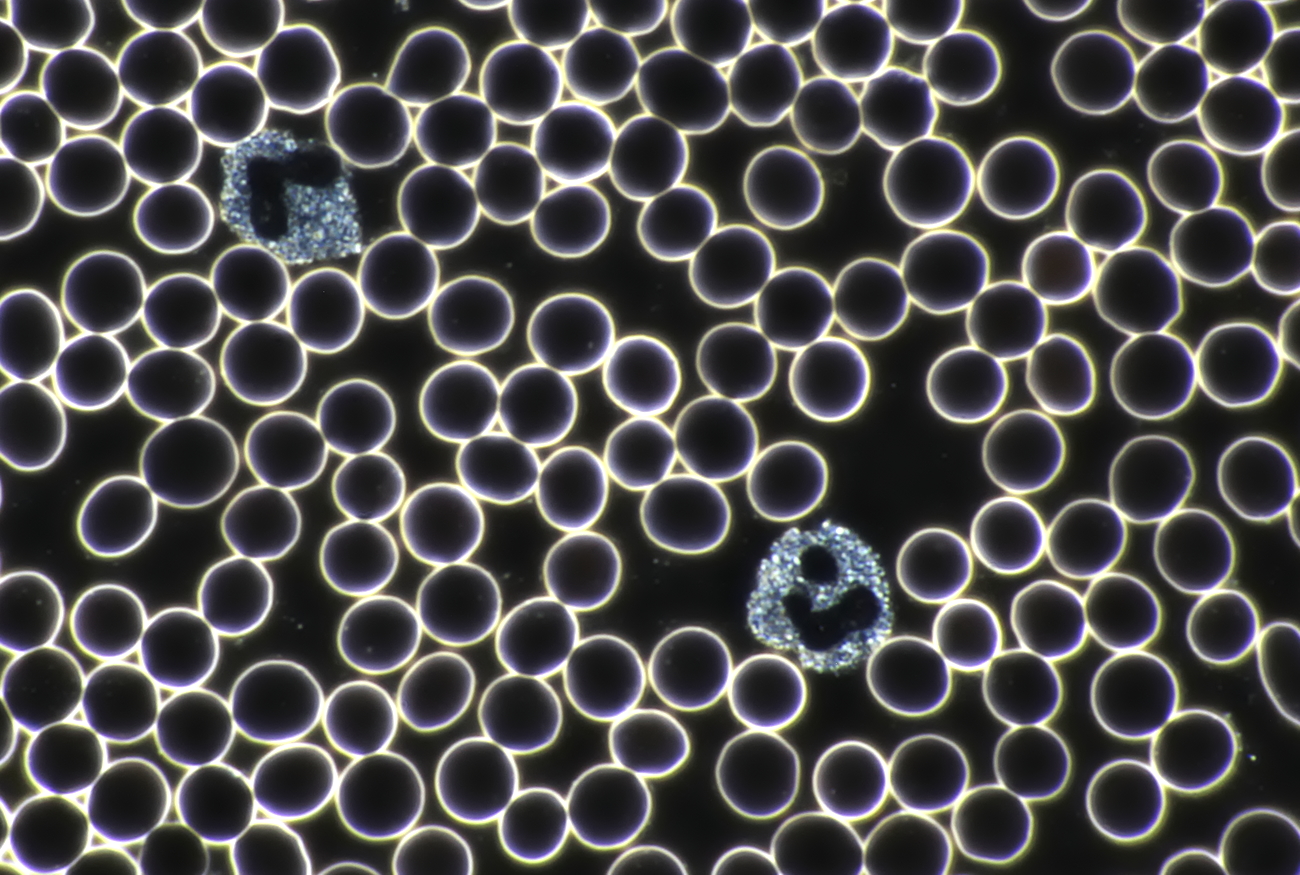
Darkfield image of blood showing two granulocytes. The nuclei and grains of leucocytes are good to be seen with darkfield illumination. Objectief: Carl Zeiss 40/1.0.
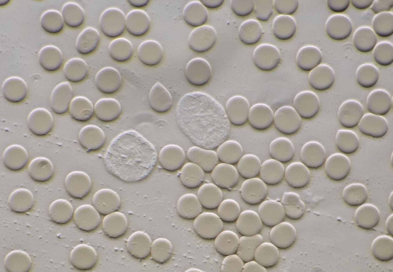
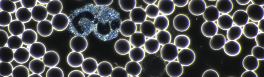
Fresh blood photographed with oblique illumination showing two leucocytes in the middle. Objective: Carl Zeiss Apo 40/1.0.
Video of fresh blood showing a moving white blood cell in the center and far right. The movements of the red blood cells are passive and are caused by currents in the slide. Objective: Carl Zeiss Apo 40/1.0.
Note:
The observation of fresh blood described here has nothing to do with the so-called live blood analysis (LBA). This method, often offered on the internet, is absolute nonsense and has no scientific basis whatsoever. Unfortunately, there are many people who allow themselves to be 'diagnosed' through LBA and pay a fair amount of money for this quackery.
Literature
Helleman, P.W., de Nooij, E.H., Overbeeke, M.A.M., Akkerman, J.W.N., Nieuwenhuis, H.K. (1987). Hematologie. Utrecht/Antwerpen: Bohn, Scheltema & Holkema.