Testate amoebae
When you think of amoebae, you probably picture transparent, slow-moving, single-celled organisms. What's less well-known, however, is that some amoebae build a shell around themselves, a kind of shelter that offers protection from predators. Within the shell (theca), there's an opening (aperture) for contact with the outside world. In some Testate amoebae, the shell is formed by secretion, while in others, it's built by assembling various particles from the surrounding environment to form a kind of masonry. Testate amoebae vary greatly in size, and some can be quite large, up to 1 mm.
Arcella
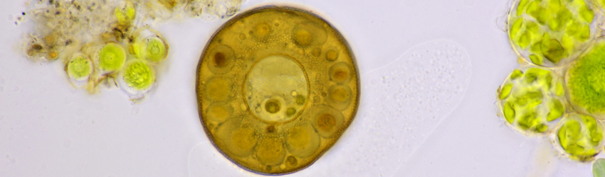
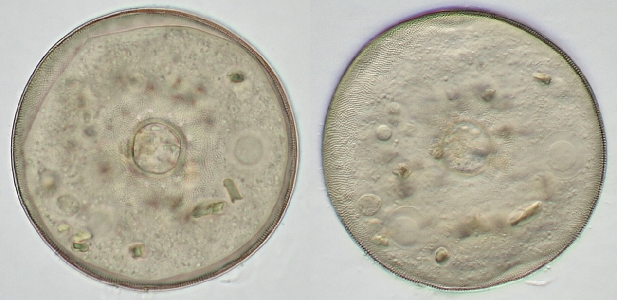
Arcella is a common testate amoeba found in fresh to brackish water that can be somewhat polluted (eutrophic). It can also be found in moist soil and mosses. Arcella's shell is sometimes quite striking. With close inspection, the amoeba can be seen within its shell. One or more nuclei may be visible; Arcella usually has two. Diatoms, algae, and other protists that the amoeba has consumed as food are also sometimes present.
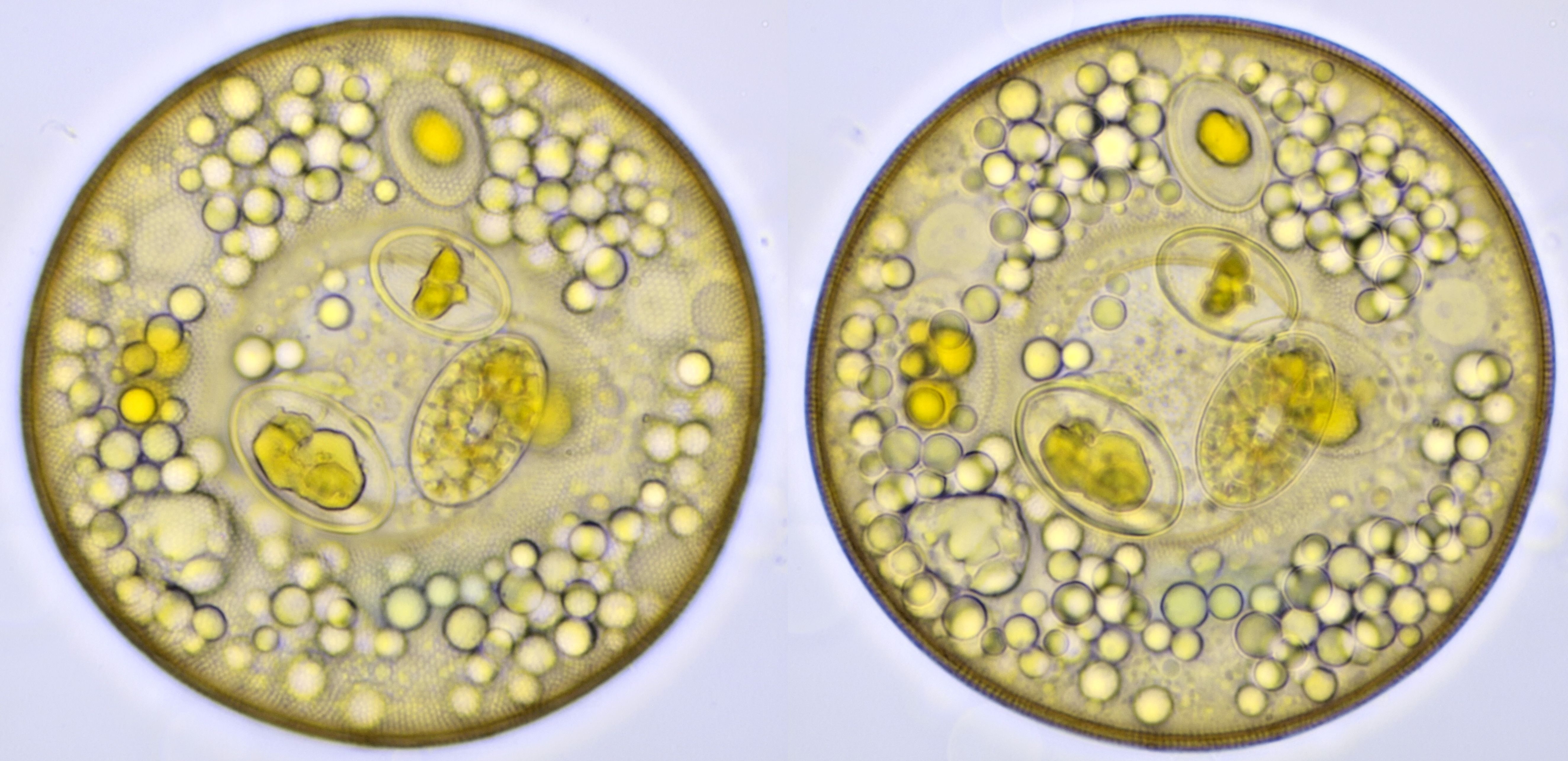
Arcella, photographed with a Carl Zeiss Neofluar 40/0.75 objective and focused on two different planes. The porous structure of the shell is visible on the left. The oval objects are diatoms ingested by the amoeba.
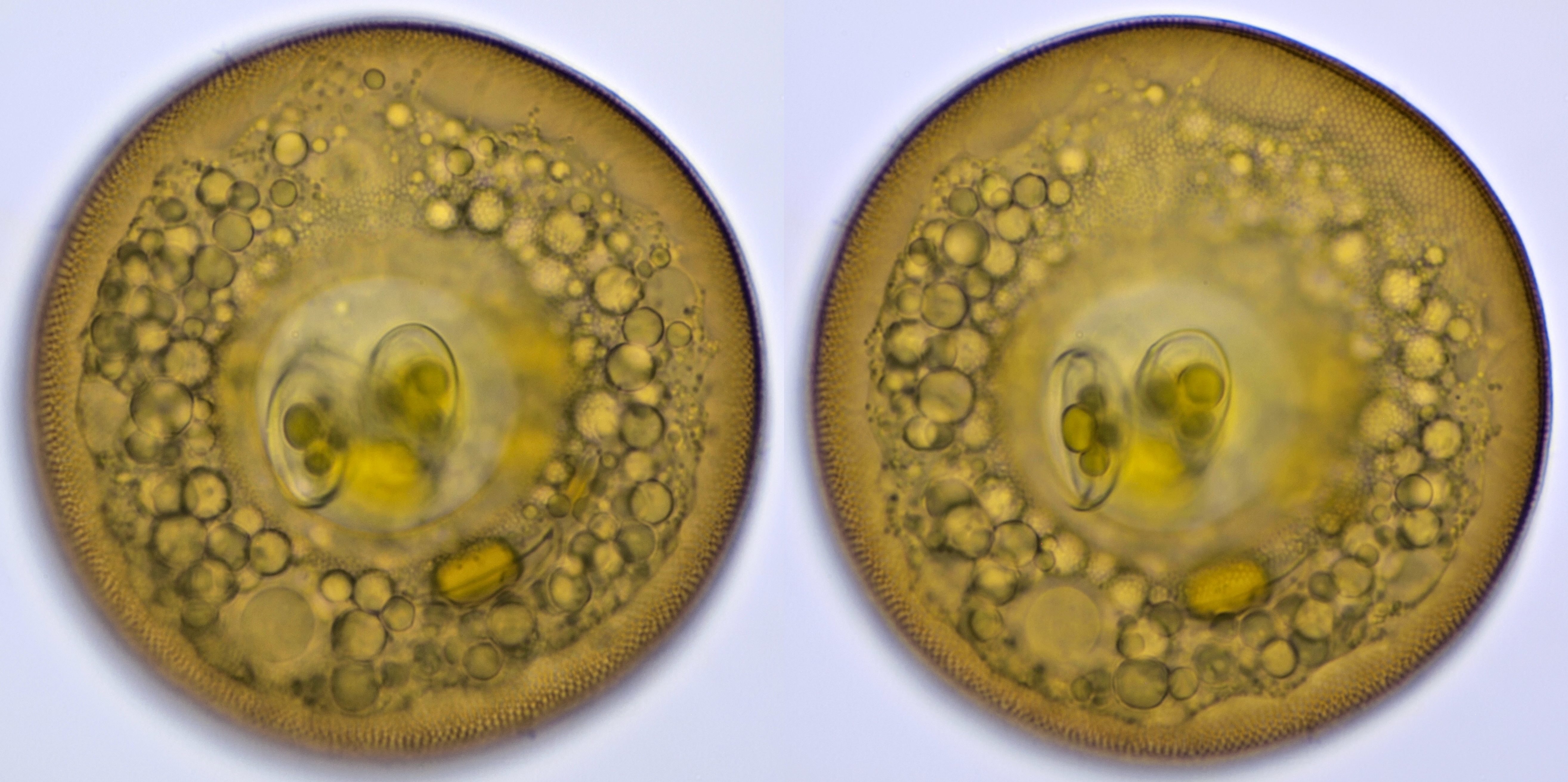
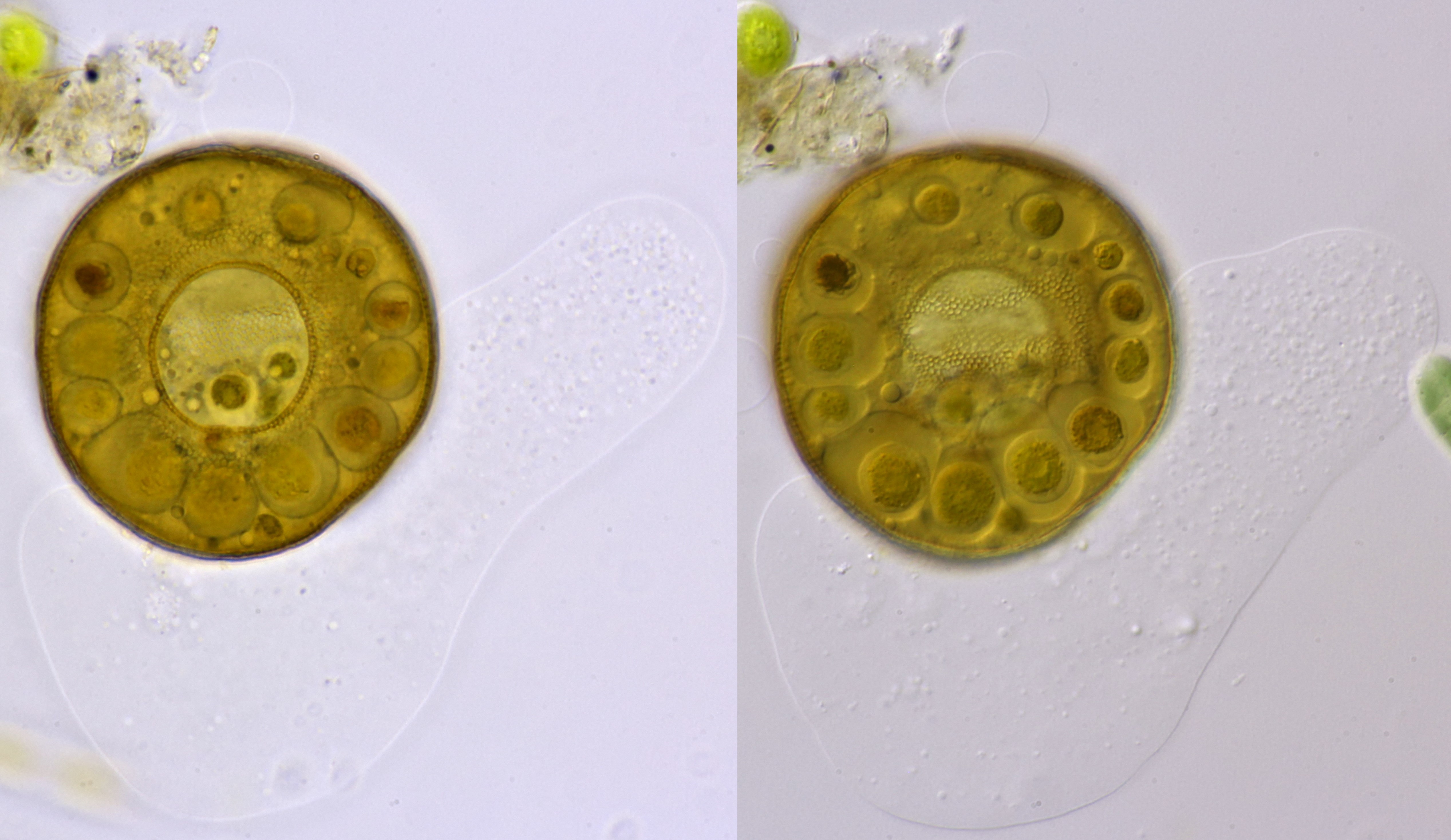
Arcella, two images of the same specimen with a slight difference in focus. Here, the contours of the amoeba are visible. A nucleus is visible at the lower left (at 7 o'clock). Objective: Carl Zeiss Neofluar 40/0.75.
Arcella from my pond. Photographed with annular illumination (left) and oblique illumination (right). A few diatoms were ingested by the amoeba. Two nuclei are present, one of which is clearly visible in the right-hand image at 11 o'clock. The annular aperture is also clearly visible in the image on the right. Objective: Carl Zeiss Apo 40/1.0
Arcella from a squeezed moss. The left image is taken in brightfield and the right image with oblique lighting. The right image again shows two nuclei. Objective: Zeiss-Winkel 40/0.65.
Video of Arcella while changing focus, showing details from the outside in.
Arcella photographed in brightfield (left) and oblique illumination (right). This specimen is infected with a parasite; these are the round objects within the shell. The amoeba is visible outside the shell and is likely dying. Objective: Carl Zeiss Apo 40/1.0.
Various Testate amoeba
A few testate amoebae that I have not identified yet are shown here.
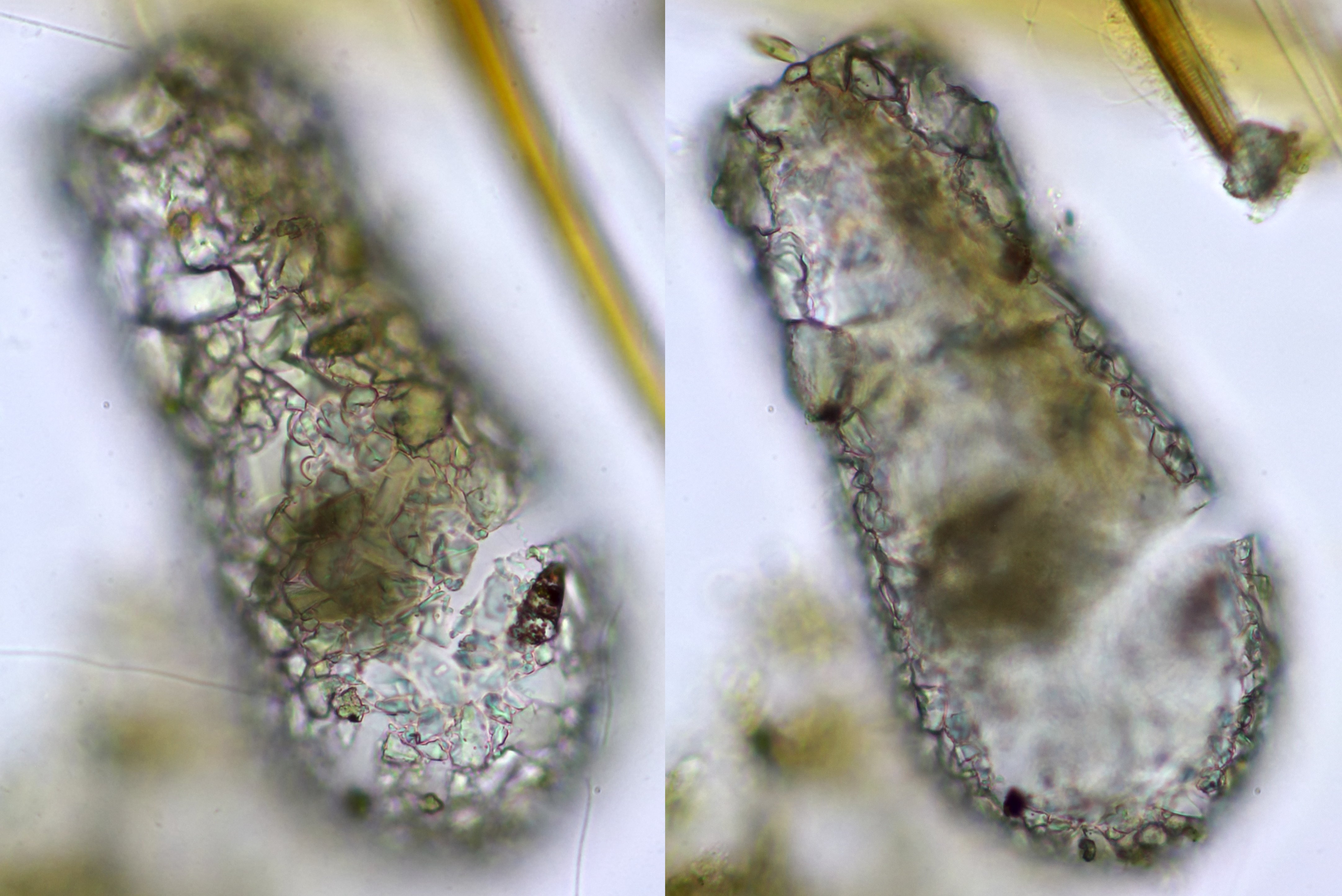
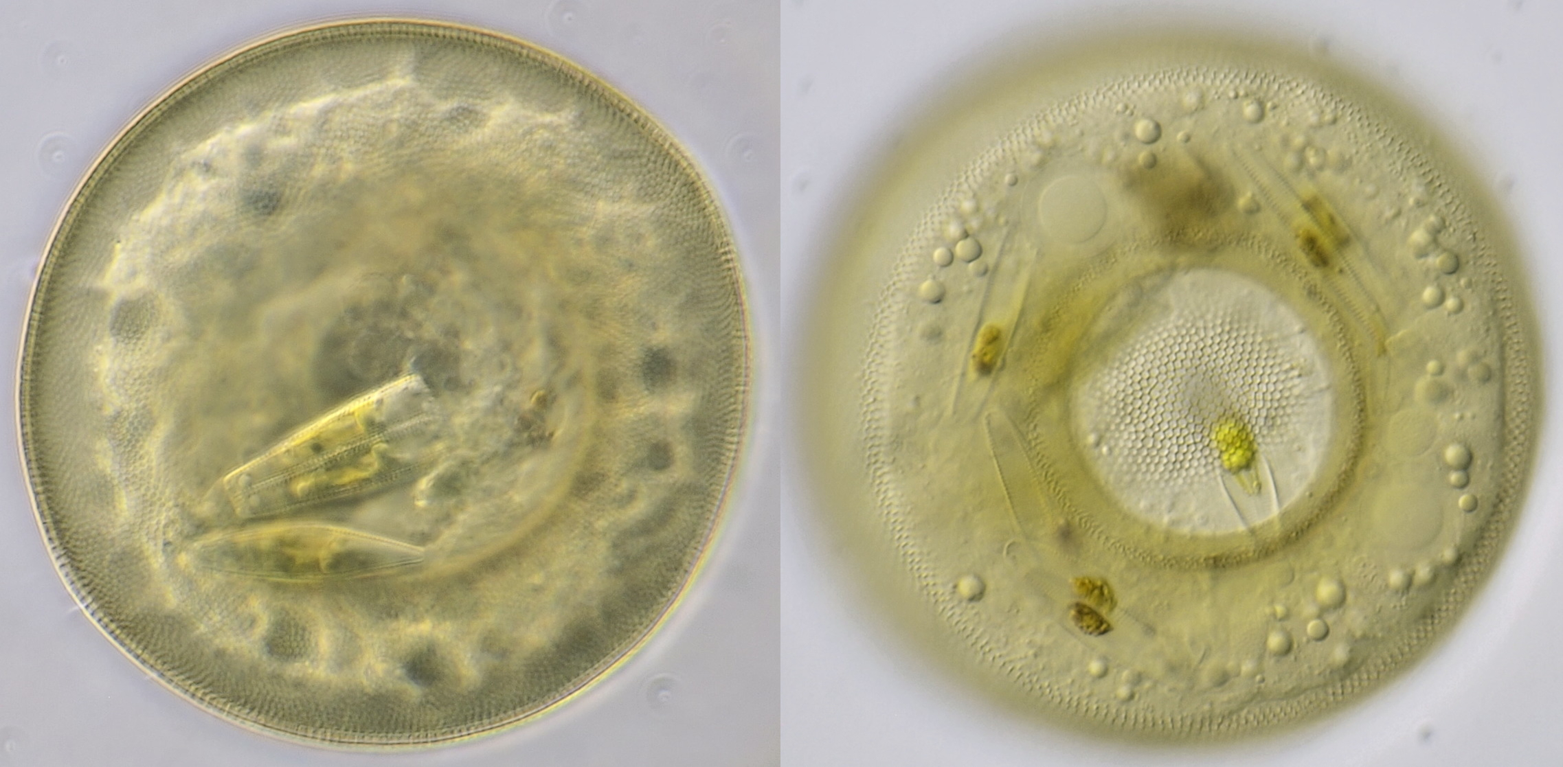
A testate amoeba, focused on two planes, clearly showing that the shell is composed of various small fragments. This specimen was found in a ditch below Duivendrecht station, the location at the church. Objective: Zeiss-Winkel 40/0.65.
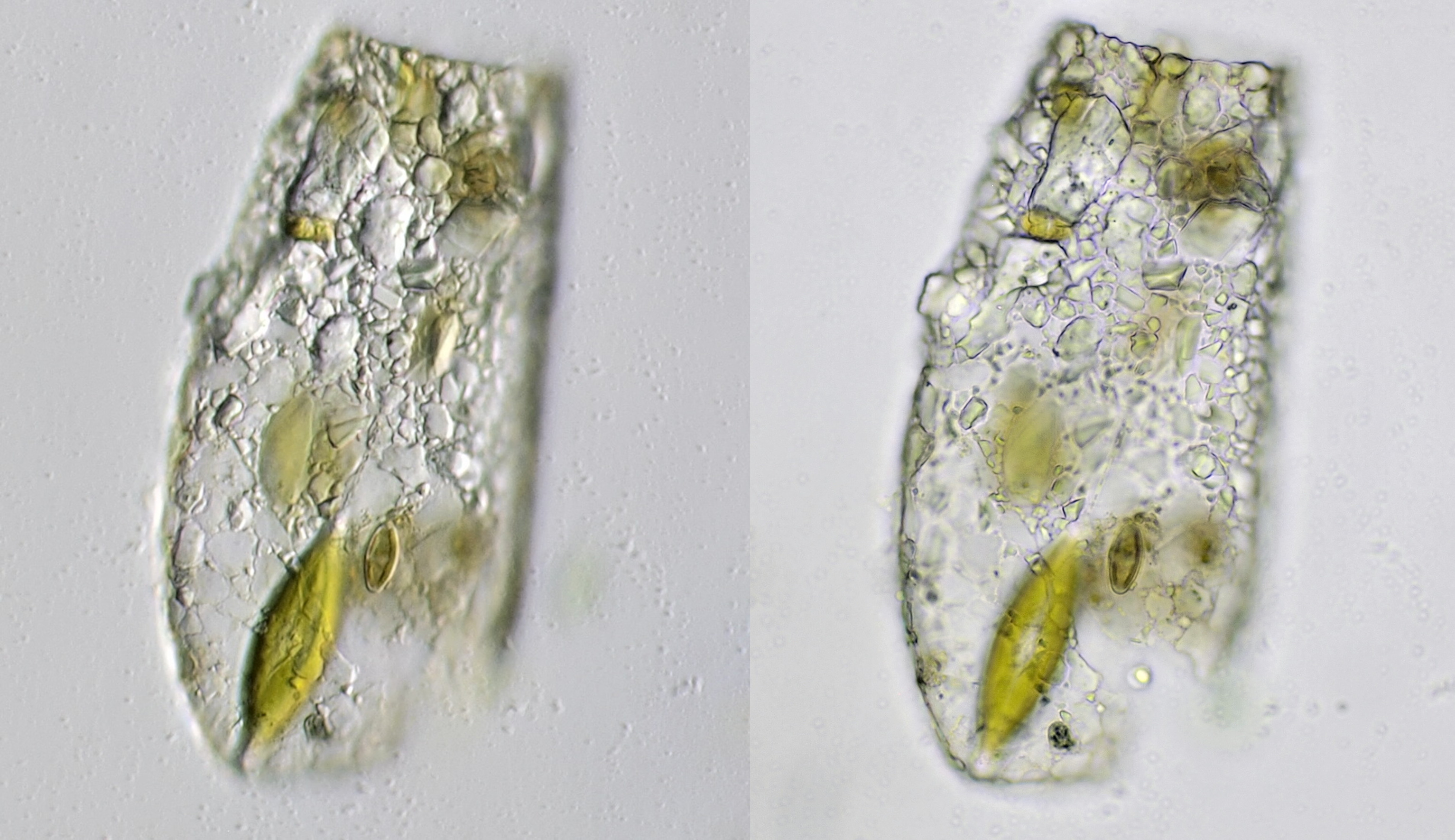
Testate amoeba photographed in oblique lighting with Leitz NPL Fluotar 25/0.55 (left) and brightfield with Leitz NPL Fluotar 40/0.70 (right).
Literature
Siemensma, Ferry (1982). Schaalamoeben (Testate amoebae). Natura.
Siemensma, F. J., Microworld, world of amoeboid organisms. World-wide electronic publication, Kortenhoef, the Netherlands.